ANAT 261 Lecture 12: Lecture 12 - Blood vessels

18 Feb 2019
School
Department
Course
Professor

October 12th, 2017
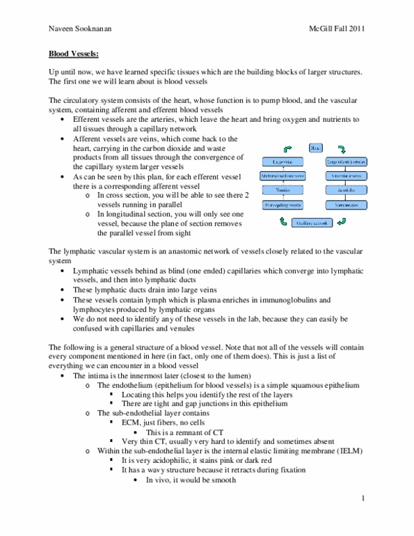
Lecture 12 – Blood Vessels
Lecture 11 – Blood Vessels Review
Elastic Arteries: Huge structures (several cm in diameter)
• Has three well-defined layers:
o (1) Intima: Innermost layer (right next to the lumen)
▪ Endothelial Layer: Simple squamous epithelium
▪ Sub-endothelial membrane: not always visible
▪ The first elastic membrane: internal elastic lamina
o (2) Media: Several successive layers of smooth muscle cells and elastic
membranes
▪ Organized
▪ Prominent → Allows elastic arteries to keep its round shape
o (3) Adventitia
Large Veins:
• Has 3 Well-defined layers:
o (1) Intima: Innermost layer (right next to the lumen)
▪ Endothelial Layer: Simple squamous epithelium
▪ Sub-endothelial membrane: not always visible
▪ Never has an IELM → Differentiates it from arteries
o (2) Media:
▪ Has a few layers of smooth muscle cells
▪ Has no elastic membranes (not organized into successive layers of
alternating elastic membranes and smooth muscle cells) →
differentiates it from arteries
▪ Collapsed structure
o (3) Adventitia: Prominent
Muscular Arteries
• Has three well-defined layers:
o (1) Intima: Innermost layer (right next to the lumen)
▪ Endothelial Layer: Simple squamous epithelium
▪ Sub-endothelial membrane: not always visible
▪ Has a visible IELM
o (2) Media: Has 4 or more layers of smooth muscle cells
▪ Thick wall of smooth muscle cells allows it to keep its shape
o (3) Adventitia: Prominent
Medium Sized Veins (or Muscular Veins)
• Has three visible layers:
o (1) Intima: Innermost layer (right next to the lumen)
▪ Endothelial Layer: Simple squamous epithelium
▪ Sub-endothelial membrane: not always visible
▪ Has no IELM (only present in efferent vessels)
o (2) Media: Has 1-2 layers of discontinuous smooth muscle cells with
adventitia penetrating it at different locations
▪ Much thinner than artery of equivalent size (muscular artery)
find more resources at oneclass.com
find more resources at oneclass.com

October 12th, 2017
▪ When the tissue is histologically prepared (formaldehyde added) →
collapses because its thin wall doesn’t support it
o (3) Adventitia: Prominent
Arteriole
• Has three well-defined layers:
o (1) Intima: Innermost layer (right next to the lumen) composed of:
▪ Endothelial Layer: Simple squamous epithelium
▪ Sub-endothelial membrane: not always visible
▪ IELM → present but not always seen
o (2) Media: count at least 2 concentric layers of smooth muscle cells
▪ If you only count 1 continuous layer, it’s probably a metarteriole, but
we’re not asked to differentiate between them in the lab so call it an
arteriole
▪ Continuous wall of smooth muscle cells allows it to keep its shape
o (3) Adventitia: Prominent
Venule
• Does not have three well-defined layers:
o Intima: Only Endothelial cells are visible and does NOT have an IELM
o Media: Not visible → structure is collapsed
o Adventitia: thin but visible
find more resources at oneclass.com
find more resources at oneclass.com

October 12th, 2017
→Cross-Section of and arteriole and venule (low mag.)
Arterioles and Venules
Higher Magnification of Arteriole and Venule in Cross-Section
• Note: Size difference between the capillaries compared to the arterioles and
venules
Arteriole
• On left side → has 1 layer of smooth muscle cells
o Some smooth muscle cells appear in cross-section (just means the
arteriole wasn’t cut in a perfect cross-section)
Venules
• May also have pericytes associated to the adventitia
• Notice in slide below that there are not smooth muscle cells but there are some
cells that resemble them called pericytes
find more resources at oneclass.com
find more resources at oneclass.com
Document Summary
October 12th, 2017: when the tissue is histologically prepared (formaldehyde added) collapses because its thin wall doesn"t support it, (3) adventitia: prominent. Arteriole: has three well-defined layers, (1) intima: innermost layer (right next to the lumen) composed of, endothelial layer: simple squamous epithelium, sub-endothelial membrane: not always visible. Ielm present but not always seen: (2) media: count at least 2 concentric layers of smooth muscle cells. Venule: does not have three well-defined layers: Intima: only endothelial cells are visible and does not have an ielm: media: not visible structure is collapsed, adventitia: thin but visible. Cross-section of and arteriole and venule (low mag. ) Higher magnification of arteriole and venule in cross-section: note: size difference between the capillaries compared to the arterioles and venules. Arteriole: on left side has 1 layer of smooth muscle cells, some smooth muscle cells appear in cross-section (just means the arteriole wasn"t cut in a perfect cross-section)