ANAT 261 Lecture 15: Lecture 15 - Digestive

18 Feb 2019
School
Department
Course
Professor

October 24th, 2017
Lecture 15 – Digestive System: Oral Cavity and Salivary Gland
Digestive System
• Consists of the digestive tract and associated glands
• Digestive Tract: Oral cavity, pharinge, esophagus, small and large intestines,
and rectum
• Associated Glands: Salivary glands, liver, and pancreas
Note: Structures in pink are those seen in this course
• Function: Obtain from the ingested food the metabolites for growth and energy
requirements.
o Before being stored or used for energy, the food must be digested and
transformed into small molecules that can be absorbed by the lining of the
digestive tract
o There will be a set of enzymes to break down the carbohydrates, lipids,
and proteins into small molecules that can be absorbed by the small
intestines
• The first step occurs in the mouth, where the food is moistened by saliva and
ground by the teeth into smaller pieces
o The saliva initiates the digestion of carbohydrates
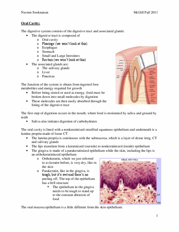
Oral Cavity
• This image is a section of a lip
Lip:
• Outside layer of lip; skin;
o Epidermis: keratinized, squamous, stratified epithelium
o Dermis: Divided into the papillary layer and the reticular layer
▪ reticular layer will be deeper, and papillary layer will be closer to the
epithelium
o Hypodermis
• As you start moving towards the mouth, there will be an area of transition where
the keratinized layer will become thinner and thinner, until it finally disappears (in
the interior of the lip, i.e. the oral mucosa)
Interior of the Lip (layers)
• Oral Mucosa: Lines the oral cavity
o Epithelial layer: non-keratinized stratified squamous epithelium
o Lamina Propria: loose connective tissue
▪ Continuous with the submucosa
• Submucosa: Denser connective tissue containing diffuse small salivary glands
• Gingiva: you will see all these indentations on both sides of the longitudinal
section of a canine tooth
o you will see a type of epithelium called parakeratinized epithelium
(Squamous, stratified epithelium that has specialized keratin called
parakeratin)
find more resources at oneclass.com
find more resources at oneclass.com

October 24th, 2017
o Para-keratin can be distinguished from ortho-keratin found on the outside
of lip
▪ Parakeratin: Wet and very hard keratin that will protect the oral
mucosa from abrasive action of some foods
• Very tough type of epithelial lining
Oral Mucosa Section
• Seen in a horizontal position (usually seen in a vertical position, like in diagram
we just looked at
• Lamina Propria: paler area
• All submucosa is just a visible collection of small salivary glands (serous type
because stain well with H&E)
o There may be some mucous glands here too
Higher Magnification of the Epithelial Covering of the Oral Mucosa
Layers of the Epithelial Covering of the Oral Mucosa:
• Base: columnar cells
• Middle: Polygonal cells (stratum spinosum)
• Upper part (facing lumen of oral cavity): Layer of squamous cells (peeling off
because it’s an epithelium in constant renewal)
Lamina Propria
• Rich capillary network
• Capillaries indicated on slide by “c”
Tongue
• Mass of striated muscle, covered by a layer of oral mucosa
• Oral Mucosa:
o Epithelium: Non-keratinized stratified squamous epithelium
o Lamina Propria: Strongly attached to the Muscular tissue
• Submucosa
o Striated Skeletal Muscle: The fibers of skeletal striated muscle run in
different directions in a section of the tongue (The skeletal muscle will be
seen in different planes of section)
Note: At the level of the oral cavity you may have some lymphoid tissue:
• Palatine Tonsil
• Lingual Tonsil: Lymphoid tissue associated to the back of the tongue
• The dorsal surface is irregular due to the presence of small eminencies called
“papillae”
• Dorsal Aspect: At the base of the tongue, there is an inverted “V”, where the
arms of that inverted “V” are towards the anterior part of the tongue
• Circumvallate Papilla: Round structures at the back of the tongue
find more resources at oneclass.com
find more resources at oneclass.com

October 24th, 2017
• Fungiform Papilla: Small round structures on dorsal aspect of tongue,
histologically different from Circumvallate Papilla
• Filiform Papilla: Makes surface of most of the dorsal aspect of the tongue rough
• At this magnification, it’s almost impossible to see the papilla
o As soon as you have a section through the tongue, you will see the
different type of papilla → Expect you to see these Papilla in lab
Papilla of the Tongue
Filiform Papilla
• Appear with a conical structure/appearance
• Tip could have a pointy or flat at top
• Rounded by squamous, stratified, non-keratinized epithelium
o Epithelium rests on a basement membrane, and under this basement
membrane you will find the lamina propria
• Could have a little keratin at their tips (Here there could be a stratum granulosum
with acidophilic granules similar to tricohyaline granules)
o Could be a little keratinized at the surface (on the tips), nowhere else on
the Filiform Papilla
Fungiform Papilla: looks like a mushroom (fungi)
• Has a narrow stalk, rounded structure
• Again, you will see this squamous, stratified, non-keratinized epithelium
o Epithelium rests on a basement membrane, and under this basement
membrane you will find the lamina propria
• May have taste buds in humans (won't be seen on animal sections on lab)
Circumvallate Papilla
• Largest of all the papilla, prominent, easy to identify
• Instead of sticking out of the mucosa, it is plunged in mucosa (not sticking out)
• Because it’s plunged into the mucosa there will be some grooves, space in
mucosa (imagine in 3D), space that goes around the papilla
• Epithelium of the oral mucosa (squamous, stratified, non-keratinized epithelium)
o Rests on a basement membrane and then almost everything underneath
the Epithelium is lamina propria
• The Epithelium gets thinner and then it’s reflected back onto the surface of the
papilla
• At the bottom of the circumvallate papilla is a collection of salivary glands (called
Von Ebner Glands)
• Von Ebner Glands:
o Serous, acini glands
o Pyramidal cells, round nucleus at the base
o Myoepithelial cells around the cells
o Pieces of the duct that open just at the bottom of the groove
find more resources at oneclass.com
find more resources at oneclass.com