NESC 2570 Lecture Notes - Lecture 6: Full Collapse, Cav2.1, Electron Microscope

27 Jun 2018
School
Department
Course
Professor

Transmitter release from synaptic vesicles
Quantal release of neurotransmitters
The neuromuscular junction as preparation to study chemical transmission
○
Miniature and plate potentials (mEPPs)
○
Relationship between mEPPs and EPPs
○
Vesicles need to fuse with the membrane to release neurotransmitters
○
○
Release of transmitter from synaptic vesicles
Ultrastructural and biochemical evidence for synaptic vesicles
○
Ultrastructural evidence that exocytosis of a single synaptic vesicle is
responsible for release of one quantum neurotransmitter
○
○
The neuromuscular junction as preparation to study chemical transmission
Neuromuscular junction used in the 1950s and 1960s to study chemical
transmission: NMJ is simple, large and easily accessible synapse
○
Motor neurons form large presynaptic terminals called end plates
○
Intracellular recording in muscle fiber near endplate. When presynaptic axon is
stimulated an excitatory postsynaptic potential, also called end plate potential
(EPP) is recorded. The EPP usually elicits action potential in the muscle
○
A very large post synaptic potential that usually reaches threshold and thereby
delivers and action potential
○
Miniature Endplate Potentials (mEPPs)
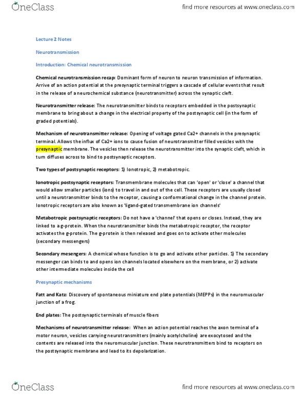
Katz and coworkers (1950s): Spontaneous changes in muscle membrane
potential occur even in the absence of motor nerve stimulation. These changes
have the same shape as EPPs but are much smaller: Miniature endplate potentials
(mEPPS)
○
The amplitude of mEPPs is rather homogenous, averaging around .5mV
○
mEPPs are too big to represent potential change in response to opening of a
single acetylcholine receptor around (.3 microvolts)
○
First Image: he got very small depolarizations of the membrane without any
stimulation. He looked at how large these miniature endplate potential were.
When he plotted them, he found they were all about the same size
It was much too big to be the opening of a single acetylcholine channel,
should be around 1000 of them opening at the same time to get a
depolarization like what he say.
○
This gave him the idea that neurotransmitter was released in quanta
(packages)
○
○
○
○
Relationship between mEPPS and EPPs
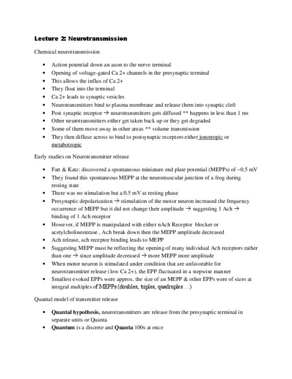
In low [Ca2+]oEPPs recorded following stimulation are very small land fluctuate
from trial to trail . Sometimes, no EPP is elicited at all (failures)
With very low calcium concentration in the bath, he stimulated a motor
neuron. Once voltage gated calcium channels opened, there was very little
calcium influx into the pre synaptic specialization and therefore there is a
very low chance release will occur. This is what he saw
○
When he stimulated this, sometimes he got no depolarization at all and
sometimes he got a very small one
○
○
The amplitude of the smallest EPPs is similar to that of mEPPs and larger EPPs
have amplitudes that are multiples of the smallest EPP sizes
○
Interpretation: EPPs are made up of individual units elicited by exocytosis of a
"quantum" of neurotransmitter, mEPPS result from the spontaneous action
potential independent release of one quantum of neurotransmitter
○
He stimulated the motor nerve in a solution with very little calcium, leaving very
little calcium influx in the post synaptic channel which results in a very little
chance of it being released
Sometimes he got no post synaptic depolarization at all, sometimes he got
larger ones
○
The smallest events were about the same size as the miniature endplates, or
they were multiples of it. This proved his hypothesis that acetylcholine was
released in packages was correct
○
If neurotransmitter is released in packages (quanta) then, what are those
packages?
○
○
Ultrastructural and biochemical evidence for synaptic vesicles
Electron microscopy of synapses
Katz' electrophysiological observations of quantal release at the NMJ
coincided with the first electron microscopy studies of synapses
○
EM studies reveal accumulations of small vesicles in presynaptic terminals
○
Katz hypothesized that neurotransmitter is stored in these vesicles and that
one quantum of neurotransmitter corresponds to the amount of transmitter
release upon exocytosis of on SV
○
When people sliced through the brain and looked at small section of the
brain
Small vesicles in large numbers which lead to believe that they
stored neurotransmitter and the physical correlator of one quanta
○
○
○
Biochemical evidence that transmitter is stored in synaptic vesicles
Synaptic vesicles biochemically isolated from brain tissue by density
gradient centrifugation; acetylcholine enriched in synaptic vesicle fractions
○
○
Vesicles might be the physical correlation of a quanta of neurotransmitter
By grinding the tissue up, we are able to isolate these vesicles
○
He would get huge amounts of acetylcholine when he did this- this is a
good indicator that these vesicles stored neurotransmitter
○
○
Ultrastructural evidence that exocytosis of a single synaptic vesicle is responsible
for release of one quantum of neurotransmitter
Heuser and Reese (1979): Correlated EM quantifications of synaptic vesicle
fusions with the quantal content (i.e. number of quanta) in EPPs
○
Used the drug 4-aminopyridine (4-AP, a potassium channel inhibitor) to increase
the number of vesicle fusion events produced by a single action potential
This drug was used for titration in this experiment
○
The action potential will not repolarize due to the potassium channel
inhibition
○
So calcium will continue coming in
○
○
Neuromuscular junction was then stimulated, rapidly frozen, and analysed using
freeze fracture electron microscopy
You freeze the specimen in a vacuum and then break the specimen.
However, because they were frozen in this state, they commonly broke
along the lipid bilayer
One lipid layer got removed while the other stayed so you are
actually looking at the inner side of the membrane
○
It is a large section of membrane - so it allows you to see much more
of the membrane
○
○
They froze it right when neurotransmitter was being released so the
neuromuscular junction would freeze immediately. They could then see the
vesicles fusing with the plasma membrane
○
○
EPPS recorded from parallel preparations to measure EPP amplitudes and
determine quantal content (number of quanta making up the EPP)
○
Number of fusing synaptic vesicles and EPP amplitude increase with increasing
concentrations of 4-AP. Agreement of estimates for number of fusing vesicles
and quantal content of EPPs
○
Exocytosis of a single synaptic vesicle leads to release of one quantum of
neurotransmitter
○
Stimulate the motor nerves and then quickly freeze the specimen - this allowed
them to catch the vesicles fusing with the plasma membrane and they correlated
the number of these events with the amount of neurotransmitter that was released
or post synaptic potential
They were able to capture the fusing of the vesicles to the membrane
○
Each hole in this image refers to a vesicle fusing with the plasma
membrane - they could correlate the fusing with the amount of
neurotransmitter released/ presynaptic potential
○
○
Freeze Fracture EM
The freeze fracture EM technique invovles breaking of frozen tissue under high
vacuum. Under these conditions, plasma membranes break between lipid layers it
fractures in a way where one lipid layer stays and the other gets removed
You're looking at large inner sections of membrane
○
○
Large expanses of the presynaptic membrane are exposed, thereby facilitating
detection of fusion synaptic vesicles. Fusing vesicles appear as pockets in the
membrane leaflets
○
Calcium in Transmitter Release
How far do we need to depolarize a membrane to create a transmitter release?
Threshold- opening of a voltage gated channel
○
○
How does an action potential in the presynaptic cell lead to the release of
neurotransmitter?
Relationship between presynaptic membrane depolarization and transmitter
release
○
The missing link between presynaptic membrane depolarization and
transmitter release: voltage gated calcium currents
○
Evidence that calcium influx through voltage gated channels is required for
transmitter release
○
Relationship between presynaptic calcium currents and transmitter release
○
○
Properties of presynaptic voltage gated calcium channels
Characteristics of calcium channel voltage activation and implications for
synaptic transmission
○
Why voltage gated calcium channels need to be in close proximity to
fusion competent synaptic vesicles to elicit release
○
Voltage gated calcium channel types involved in transmitter release
○
Diseases associated with presynaptic calcium channels
○
○
Relationship between presynaptic membrane depolarization and transmitter
release
Squid giant synapse preparation: Allow simultaneous voltage-clamp of the
presynaptic terminal and intracellular recording of the membrane potential in the
postsynaptic cell (3 electrodes needed)
Voltage gated sodium and voltage gated potassium channels must be
blocked in order to determine the dependency of membrane depolarization.
So action potential must be restricted
○
○
Katz and Miledi (1967): Voltage-gated sodium channels blocked with
tetrodotoxin. Neurotransmitter release elicited by injecting current into the
presynaptic terminal and recording from the post synaptic neuron
Electron clamping allows the systematic increase of depolarization and see
how the pre synaptic membrane would change and would see if he got a
post synaptic action potential or not
○
Result: if he injected a large depolarization in the pre synaptic neuron, he
would only get a slight depolarization in the post synaptic neuron. If he
injected a slightly larger depolarization, then he would get a larger
depolarization at the post synaptic membrane etc.
○
The experiment tells us that we need to open a voltage gated channel and
the pre synaptic membrane and that causes neurotransmitter release
○
Caused by voltage gated calcium channels
○
○
Postsynaptic potential shows steep dependence on presynaptic depolarization:
With depolarizations of 50 mV or less (presynaptic potential -20 mV or less), no
EPSP; with depolarizations of 70 mV or more, maximal EPSP.
○
When he injected a depolarizing current, he would assess whether or not he got a
post synaptic potential
If he injected a small depolarizing current, he would not get a post synaptic
potential
○
○
This experiment tells us that we need to open a voltage gated channel in the pre
synaptic membrane
These must be voltage gated calcium channels to cause neurotransmitter
release
○
So we can correlate voltage gated calcium channels (currents) with the post
synaptic potential.
○
○
The missing link between presynaptic membrane depolarization and transmitter
release: Voltage-gated calcium currents
Voltage-dependent calcium currents (ICa) can be detected by voltage clamping
presynaptic terminals (e.g. in the squid giant synapse preparation). Blockade of
voltage-dependent sodium and potassium currents with tetrodotoxin and
tetraethylammonium, respectively, isolates ICa.
○
The amplitude of the calcium currents correlates well with the amplitudes of
postsynaptic currents
○
○
Evidence that voltage gated calcium currents are required for transmitter release
Buffering of intracellular calcium with a fast calcium chelator abolishes synaptic
transmission
○
Calcium that is bound to a chelator can no longer bind to the receptors and elicit
a release
You will have a presynaptic potential but no post synaptic potential
○
○
You can do the inverse, if you inject calcium in the pre synaptic terminal ,you
will elicit a post synaptic depolarization
Calcium is necessary and sufficient to elicit release
○
○
Injection of calcium into the presynaptic terminal leads to a postsynaptic
potential in the absence of presynaptic membrane depolarization
○
Increasing the extracellular calcium concentration increases the amplitude of
postsynaptic currents, whereas decreasing [Ca2+] o attenuates and ultimately
abolishes synaptic transmission 3.
○
You can vary the extra cellular calcium concentration if you have a high
extracellular concentration, you will have a large pre synaptic potential. If you
drop the extra cellular concentration, you will drop the potential
As you change calcium concentration, the amount of calcium entering the
cell will change and therefore alter the action potential
○
○
Blockade of voltage-gated calcium channels abolishes synaptic transmission
People used cadmium to do this to block the channels
○
This abolished the post synaptic response, proving that calcium is
necessary for depolarization in the post synaptic neuron
○
○
Relationship between presynaptic voltage gated calcium currents and transmitter
release
The postsynaptic current (and potential) shows steep dependence on presynaptic
calcium current:
Linear relationship - if you increase your pre synaptic calcium
concentration, you get a significantly larger post synaptic current
○
Small changes in calcium lead to large changes in neurotransmitter release
○
○
When you plot the presynaptic current to the post synaptic current, you have a
linear relationship
Meaning that if you increase the presynapic calcium current you get a
much larger post synaptic current
○
Post synaptic current vs calcium current is that post synaptic current
associates with the third or fourth power of calcium
○
○
The calcium sensor triggering neurotransmitter release must bind 3-4 calcium
ions in a cooperative manner
Proteins have low affinity to the first calcium ion, once this ion has bound,
it makes it much more easier for the second ion to bind
○
Reason why the changes in the calcium influx leads to large changes in
release
○
○
Characteristics of calcium channel voltage activation and implications for
synaptic transmission
There is a long delay between the action potential (sodium current) and the post
synaptic current
This is because the neurotransmitter has to diffuse across the synaptic cleft,
but this only takes 100's of miliseconds
○
Calcium channels actually open very slowly AFTER the depolarization
○
Membrane depolarizes and it takes between 1 and 2 miliseconds for
voltage gated calcium channels to open
○
○
Voltage-dependent calcium channels, in comparison to sodium or potassium
channels, activate only slowly in response to membrane depolarization. As a
consequence, ICa is delayed by about 0.8 ms relative to the action potential.
○
The delayed opening of VGCCs accounts for much of the synaptic delay. (In
comparision, diffusion of neurotransmitter across the synaptic cleft only accounts
for a delay of 0.2 ms).
○
Why presynaptic voltage gated calcium channels need to be in close proximity to
synaptic vesicles
Calcium is effectively buffered in all eukaryotic cells, cocnentration of calcium
inside cells is about 100 nm
Calcium is a very important second messanger so we want t keep the
concentration fairly low so we can elicit signalling
○
All eukaryotic cells have evolved mechanisms and a lot of calcium binding
proteins whose main function is to bind calcium and take it out of the
equation
○
This is also true for neurons - if a voltage gated calcium channel opens, the
calcium concentration will rise very fast and the mouth of the channel and
will alter the gradient of calcium in the cytosol
○
○
While the extracellular calcium concentration is high (10-3 M range), the
intracellular concentration is very low (10-7 M).
○
Besides having efficient calcium extrusion mechanisms (calcium pumps in
plasma membrane) and intracellular stores for calcium, neurons keep the free
intracellular calcium concentration low by expressing calcium-binding proteins.
○
If voltage-dependent calcium channels open, inflowing calcium is quickly
buffered by calcium-binding proteins, creating a steep calcium gradient at the
channel mouth
○
The calcium sensor responsible for triggering synaptic vesicle fusion has a fairly
low affinity to calcium (Kd between 10-5 and 10-4 M) and therefore has to be
very close to an open calcium channel: Only synaptic vesicles present in a
calcium micro- or nanodomain around open calcium channels can undergo
calcium-triggered fusion
○
The calcium sensory for neurotransmitter release has a very low affinity for
calcium
So calcium has to be very close to the mouth of a channel to fuse and allow
for neurotransmitter release
○
synaptic vesicles
○
○
Voltage Gated Calcium Channel Types Involved in Transmitter release
Type Alpha 1
Subunit
Activation
Inhibitors
Function in transmitter release
P/Q Alpha 1A
(Cav2.1)
HVA(1) omega
agatoxin
Fast transmitter release
N Alpha 1B
(Cav2.2)
HVA(1) omega
conotoxin
Fast transmitter release
R Alpha 1E
(CaV2.3)
HVA(1) Minor role in fast release; Ca2+ dependent
plasticity of release
L Alpha 1C,D,F,S
(CaV1.1-1.4)
HVA(1)
dihydrophyrid
ines
Transmitter release to graded
depolarizations in some sensory neurons;
slow release of peptide transmitters
T Alpha 1G,H,I
(CaV3.1-3.3)
LVA(2) Transmitter release in response to graded
depolarizations in some sensory neurons
(?)
Note: HVA I High Voltage Activation - activate positive to -30 to -20 mV
Mediate most of the fast neurotransmitter release - neurotransmitters that
elicit an immediate response at the post synaptic terminal such as GABA
○
○
Note 2.0: LVA means low voltage activation - activate positive to -70mV
Open if the membrane is only slightly depolarized
○
○
Predominance of P/Q and N-type VGCCs in fast transmitter release
○
This predominance is due to preferential localization of P/Q and N type VFCCs
to release sites: Protein interactions with components of the exocytosis
machinery
○
Other VGCC types (such as L-type channels) may be involved in slow release of
dense core vesicles (peptide neurotransmitter) and in transmitter release in
response to graded depolarization (ex. Photoreceptors)
○
Very specific inhibitors to these calcium channels that occur in the animal
Ex. Sea slugs have a toxin that they inject into fish that ceases the
neuromuscular junctions from working (and calcium channels)
○
○
Diseases Associated with Presynaptic Calcium Channels
Mutations in P/Q type calcium channels leading to congenital diseases
Mutations in P/Q type calcium channels cause a variety of congenital
syndromes: Familial Hemiplegic Migraine certain ataxias (inability to
coordinate voluntaria movements, often characteristics of cerebellar or
spinal cord defects), and absence epilepsy (seizures characterised by
unconsciousness, usually without involuntary muscle contractions)
○
No disease causing mutations in N type calcium channels known
○
Lead to migrains
○
Lead to difficulty in controlling motor functions
○
○
Lambert- Eaton myastenic syndrome (LEMS)
Compliations in patients ith certain kinds of cancers, especially small cell
lung carcioma
○
Weakness and fatigability of skeletal muscles
○
Electrophysiology on muscle biopsies: Quantal content of Epps greatly
reduced; mEPP amplitude unchanged. Raising the extracellular calcium
concentration increases EPP amplitude
○
Histology: Lower density of calcium channels
○
Autoimmune disease: Blood of LEMS patients contains high
concentrations of antibodies to presynaptic calcium channels
○
Antibodies bind to calcium receptors and if this happens, you have little
control over your muscles because calcium cannot bind
○
○
Biogenesis and Local Recycling of Synaptic Vesicles
Biogenesis of synaptic vesicles containing small-molecule neurotransmitters
○
Biogenesis of synaptic vesicles containing peptide neurotransmitters
○
Evidence for local recycling of synaptic vesicles
○
The synaptic vesicle cycle
○
Different routes for synaptic vesicle exocytosis?
○
Synaptic vesicle pools
○
After synaptic vesicles fuse with the plasma membrane…
Depends on the type of neurotransmitter
○
Vesicles are created locally at the synapse
○
Once they fuse with the membrane, they actually recycle
○
○
Biogenesis of Synaptic Vesicles Containing Small Molecule Neurotransmitters
Synthesis and/or uptake of small molecules neurotransmitters occurs locally
within presynaptic terminals
Some neurotransmitters (such as glutamate) are taken up from the
extracellular space by plasma membrane transporters
○
For some neurotransmitters, precursors are taken up from the extracellular
space. Enzymes produced in the soma and transported to the terminal via
slow axonal transport, locally synthesize the neurotransmitter from its
precursor
○
Neurotransmitter is loaded into synaptic vesicles by a vesicular transporter
○
○
Loading of Small Molecule Transmitters into Synaptic Vesicles
Neurotransmitters are loaded into synaptic vesicles against a concentration
gradient by vesicular neurotransmitter transporters
○
Energy for this transport comes from an electrochemical gradient across the
synaptic vesicle membrane that is created by the vesicular proton pump (V type
H+-ATPase)
○
The vesicular proton pump hydrolyzes ATP to transport protons into the synaptic
vesicle lumen, creating a pH gradient and membrane potential (Δψ)
○
The NT transporters use a proton antiport, the membrane potential or both to
translocate neurotransmitter into the vesicle lumen
○
Neurotransmitter is loaded into the synaptic vesicle
Concentration is larger inside the vesicle than the outside
○
So lots of energy is needed to move the neurotransmitter against its
concentration gradient
○
This process, all of the time, transporter is used that allows for secondary
active transport
The transporter co transports a second compound that is moved
WITH the concentration gradient
○
It uses one electrochemical gradient to move the neurotransmitter
against its electrochemical gradient
○
Second compound that is being transported is proteins
○
○
The lumen is much more acidic than the cytosol because the vesicles
contain a proton pump
○
These pump utilize ATP to pump protons into the synaptic vesicle
○
Protons is used to transport neurotransmitter into the vesicle
○
○
Biogenesis of Synaptic Vesicles Containing Peptide Neurotransmitters
Neuropeptides are synthesized in the soma (ER to the golgi)
Ribosomes in the ER are converted into neuropeptides in the golgi and
then are transported all the way to the synapse
○
This is much different than how fast neurotransmitter is loaded
○
○
Peptide filled large dense core vesicles are transported along microtubules via
fast axonal transport (up to 5 micrometers/second)
○
Neuropeptides do not undergo re-uptake; rather, they are degraded by proteolytic
enzymes
○
Membrane on the synaptic vesicle is recycled
○
Evidence for Local Recycling of Synaptic Vesicles
Synaptic vesicule fusion adds new membrane to the plasma membrane. Plasma
membrane surface area usually held constant by compensatory endcytosis
○
Evidence Horseradish peroxidase (HRP: an enzyme that can be made to produce
an electron dense reaction product) applied extracellularly to a neuromuscular
junction preparation
○
Repetitive stimulation of afferent nerve leads to uptake of HRP into the nerve
terminal
○
Preparation fixed and processed for electron microscopy at different times
following stimulation: HRP first seen in coated pits then in endosome like
vesicles, finally in synaptic vesicles -> Synaptic vesicles recycle
○
Can put a type of dye in the bath where you have neurons and then trigger
activation of neurons
When they are exocytose, they endocytose the membrane and the dye and
view it in the pre synaptic terminal
○
○
The Synaptic Vesicle Cycle
Observations from early HRP studies and recent styrl dye experiments indicate:
Vesicular membrane is retrieved by clathrin mediated endocytosis which is
completed 10-20 seconds following exocytosis
○
The majority of all endocytosed vesicles likely bypasses endosomes,
immediately becoming synaptic vesicles after uncoating
○
Synaptic vesicles have to dock to the active zone and undergo a priming
step before becoming fusion competent. A synaptic vesicle can complete
the whole endocytosis cycle in approximately 1 minute
○
It takes about 10-20 seconds to endocytose the membrane, the newly
formed vesicles are then available to be released
○
In many membranes, you only have about 100 vesicles, this means that if
you need a minute for each synaptic vesicle to release, you can use up the
vesicles in the pool 100 or several hundred times, you will have no vesicles
yet
However synapses must fire more often than that
○
Sometimes, synaptic vesicles don't fully fuse with the membrane - so
it is immediately available for another release
○
○
○
Different Routes for Synaptic Vesicle Exocytosis?
Experiments employing styryl dyes as well as studies using capacitance
measurements have led to the suggestion that two different exocytosis
mechanisms may coexist:
"Classical" Exocytosis
Full collapse of vesicle membrane into plasma membrane
○
Clathrin-mediated endocytosis to retrieve synaptic vesicle
membranes requires 20 seconds
○
High frequency stimulation quickly leads to deletion of vesicles at
synapses with relatively small synaptic vesicle pools
○
○
"Kiss-and-run" exocytosis
Transient fusion pore; vesicle membrane never fully collapses into
plasma membrane
○
Possibly repeated fusions of individual synaptic vesicle with plasma
membrane in short time frame
○
May allow for sustained release in response to repetitive stimulation
○
○
○
Synaptic Vesicle Pools
Not all synaptic vesicles at a given release site can undergo exocytosis. Studies
employing styryl dyes as well as electrophysiological experiments allow to
distinguish several functionally different pools of synaptic vesicles:
Readily releasable pool of synaptic vesicles
Pool of synaptic vesicles immediately available for release
○
At CNS synapses, only 2-4% of all synaptic vesicles ( 5-10 vesicles)
16 synaptic vesicles that can be recruited to release the content
if there is more than one action potential
○
About 80 don't do shit - this is because they don't contain all
the proteins required undergo fusion with the membrane.
○
Synapses have to be VERY economical in how they release
neurotransmitter and how they use vesicles
○
○
Readily releasable vesicles may correspond to synaptic vesicles
docked to the active zone
○
○
"Reserve pool" of synaptic vesicles
Synaptic vesicles available for exocytosis but not for immediate
release
○
Together, readily releasable and reserve pool constitute the recycling
pool of synaptic vesicles, representing on average 20% of all
synaptic vesicles
○
○
"Resting pool" of synaptic vesicles
Non recycling synaptic vesicles; largest pool
○
○
How many synaptic vesicles actually recycle? Only about 20% of all
synaptic vesicles are available for neurotransmitter release
Part of this reserved pool of synaptic vesicles
○
○
○
Neurotransmitter Release
October 21, 2015
1:32 PM

Transmitter release from synaptic vesicles
Quantal release of neurotransmitters
The neuromuscular junction as preparation to study chemical transmission
○
Miniature and plate potentials (mEPPs)
○
Relationship between mEPPs and EPPs
○
Vesicles need to fuse with the membrane to release neurotransmitters
○
○
Release of transmitter from synaptic vesicles
Ultrastructural and biochemical evidence for synaptic vesicles
○
Ultrastructural evidence that exocytosis of a single synaptic vesicle is
responsible for release of one quantum neurotransmitter
○
○
The neuromuscular junction as preparation to study chemical transmission
Neuromuscular junction used in the 1950s and 1960s to study chemical
transmission: NMJ is simple, large and easily accessible synapse
○
Motor neurons form large presynaptic terminals called end plates
○
Intracellular recording in muscle fiber near endplate. When presynaptic axon is
stimulated an excitatory postsynaptic potential, also called end plate potential
(EPP) is recorded. The EPP usually elicits action potential in the muscle
○
A very large post synaptic potential that usually reaches threshold and thereby
delivers and action potential
○
Miniature Endplate Potentials (mEPPs)
Katz and coworkers (1950s): Spontaneous changes in muscle membrane
potential occur even in the absence of motor nerve stimulation. These changes
have the same shape as EPPs but are much smaller: Miniature endplate potentials
(mEPPS)
○
The amplitude of mEPPs is rather homogenous, averaging around .5mV
○
mEPPs are too big to represent potential change in response to opening of a
single acetylcholine receptor around (.3 microvolts)
○
First Image: he got very small depolarizations of the membrane without any
stimulation. He looked at how large these miniature endplate potential were.
When he plotted them, he found they were all about the same size
It was much too big to be the opening of a single acetylcholine channel,
should be around 1000 of them opening at the same time to get a
depolarization like what he say.
○
This gave him the idea that neurotransmitter was released in quanta
(packages)
○
○
○
○
Relationship between mEPPS and EPPs
In low [Ca2+]oEPPs recorded following stimulation are very small land fluctuate
from trial to trail . Sometimes, no EPP is elicited at all (failures)
With very low calcium concentration in the bath, he stimulated a motor
neuron. Once voltage gated calcium channels opened, there was very little
calcium influx into the pre synaptic specialization and therefore there is a
very low chance release will occur. This is what he saw
○
When he stimulated this, sometimes he got no depolarization at all and
sometimes he got a very small one
○
○
The amplitude of the smallest EPPs is similar to that of mEPPs and larger EPPs
have amplitudes that are multiples of the smallest EPP sizes
○
Interpretation: EPPs are made up of individual units elicited by exocytosis of a
"quantum" of neurotransmitter, mEPPS result from the spontaneous action
potential independent release of one quantum of neurotransmitter
○
He stimulated the motor nerve in a solution with very little calcium, leaving very
little calcium influx in the post synaptic channel which results in a very little
chance of it being released
Sometimes he got no post synaptic depolarization at all, sometimes he got
larger ones
○
The smallest events were about the same size as the miniature endplates, or
they were multiples of it. This proved his hypothesis that acetylcholine was
released in packages was correct
○
If neurotransmitter is released in packages (quanta) then, what are those
packages?
○
○
Ultrastructural and biochemical evidence for synaptic vesicles
Electron microscopy of synapses
Katz' electrophysiological observations of quantal release at the NMJ
coincided with the first electron microscopy studies of synapses
○
EM studies reveal accumulations of small vesicles in presynaptic terminals
○
Katz hypothesized that neurotransmitter is stored in these vesicles and that
one quantum of neurotransmitter corresponds to the amount of transmitter
release upon exocytosis of on SV
○
When people sliced through the brain and looked at small section of the
brain
Small vesicles in large numbers which lead to believe that they
stored neurotransmitter and the physical correlator of one quanta
○
○
○
Biochemical evidence that transmitter is stored in synaptic vesicles
Synaptic vesicles biochemically isolated from brain tissue by density
gradient centrifugation; acetylcholine enriched in synaptic vesicle fractions
○
○
Vesicles might be the physical correlation of a quanta of neurotransmitter
By grinding the tissue up, we are able to isolate these vesicles
○
He would get huge amounts of acetylcholine when he did this- this is a
good indicator that these vesicles stored neurotransmitter
○
○
Ultrastructural evidence that exocytosis of a single synaptic vesicle is responsible
for release of one quantum of neurotransmitter
Heuser and Reese (1979): Correlated EM quantifications of synaptic vesicle
fusions with the quantal content (i.e. number of quanta) in EPPs
○
Used the drug 4-aminopyridine (4-AP, a potassium channel inhibitor) to increase
the number of vesicle fusion events produced by a single action potential
This drug was used for titration in this experiment
○
The action potential will not repolarize due to the potassium channel
inhibition
○
So calcium will continue coming in
○
○
Neuromuscular junction was then stimulated, rapidly frozen, and analysed using
freeze fracture electron microscopy
You freeze the specimen in a vacuum and then break the specimen.
However, because they were frozen in this state, they commonly broke
along the lipid bilayer
One lipid layer got removed while the other stayed so you are
actually looking at the inner side of the membrane
○
It is a large section of membrane - so it allows you to see much more
of the membrane
○
○
They froze it right when neurotransmitter was being released so the
neuromuscular junction would freeze immediately. They could then see the
vesicles fusing with the plasma membrane
○
○
EPPS recorded from parallel preparations to measure EPP amplitudes and
determine quantal content (number of quanta making up the EPP)
○
Number of fusing synaptic vesicles and EPP amplitude increase with increasing
concentrations of 4-AP. Agreement of estimates for number of fusing vesicles
and quantal content of EPPs
○
Exocytosis of a single synaptic vesicle leads to release of one quantum of
neurotransmitter
○
Stimulate the motor nerves and then quickly freeze the specimen - this allowed
them to catch the vesicles fusing with the plasma membrane and they correlated
the number of these events with the amount of neurotransmitter that was released
or post synaptic potential
They were able to capture the fusing of the vesicles to the membrane
○
Each hole in this image refers to a vesicle fusing with the plasma
membrane - they could correlate the fusing with the amount of
neurotransmitter released/ presynaptic potential
○
○
Freeze Fracture EM
The freeze fracture EM technique invovles breaking of frozen tissue under high
vacuum. Under these conditions, plasma membranes break between lipid layers it
fractures in a way where one lipid layer stays and the other gets removed
You're looking at large inner sections of membrane
○
○
Large expanses of the presynaptic membrane are exposed, thereby facilitating
detection of fusion synaptic vesicles. Fusing vesicles appear as pockets in the
membrane leaflets
○
Calcium in Transmitter Release
How far do we need to depolarize a membrane to create a transmitter release?
Threshold- opening of a voltage gated channel
○
○
How does an action potential in the presynaptic cell lead to the release of
neurotransmitter?
Relationship between presynaptic membrane depolarization and transmitter
release
○
The missing link between presynaptic membrane depolarization and
transmitter release: voltage gated calcium currents
○
Evidence that calcium influx through voltage gated channels is required for
transmitter release
○
Relationship between presynaptic calcium currents and transmitter release
○
○
Properties of presynaptic voltage gated calcium channels
Characteristics of calcium channel voltage activation and implications for
synaptic transmission
○
Why voltage gated calcium channels need to be in close proximity to
fusion competent synaptic vesicles to elicit release
○
Voltage gated calcium channel types involved in transmitter release
○
Diseases associated with presynaptic calcium channels
○
○
Relationship between presynaptic membrane depolarization and transmitter
release
Squid giant synapse preparation: Allow simultaneous voltage-clamp of the
presynaptic terminal and intracellular recording of the membrane potential in the
postsynaptic cell (3 electrodes needed)
Voltage gated sodium and voltage gated potassium channels must be
blocked in order to determine the dependency of membrane depolarization.
So action potential must be restricted
○
○
Katz and Miledi (1967): Voltage-gated sodium channels blocked with
tetrodotoxin. Neurotransmitter release elicited by injecting current into the
presynaptic terminal and recording from the post synaptic neuron
Electron clamping allows the systematic increase of depolarization and see
how the pre synaptic membrane would change and would see if he got a
post synaptic action potential or not
○
Result: if he injected a large depolarization in the pre synaptic neuron, he
would only get a slight depolarization in the post synaptic neuron. If he
injected a slightly larger depolarization, then he would get a larger
depolarization at the post synaptic membrane etc.
○
The experiment tells us that we need to open a voltage gated channel and
the pre synaptic membrane and that causes neurotransmitter release
○
Caused by voltage gated calcium channels
○
○
Postsynaptic potential shows steep dependence on presynaptic depolarization:
With depolarizations of 50 mV or less (presynaptic potential -20 mV or less), no
EPSP; with depolarizations of 70 mV or more, maximal EPSP.
○
When he injected a depolarizing current, he would assess whether or not he got a
post synaptic potential
If he injected a small depolarizing current, he would not get a post synaptic
potential
○
○
This experiment tells us that we need to open a voltage gated channel in the pre
synaptic membrane
These must be voltage gated calcium channels to cause neurotransmitter
release
○
So we can correlate voltage gated calcium channels (currents) with the post
synaptic potential.
○
○
The missing link between presynaptic membrane depolarization and transmitter
release: Voltage-gated calcium currents
Voltage-dependent calcium currents (ICa) can be detected by voltage clamping
presynaptic terminals (e.g. in the squid giant synapse preparation). Blockade of
voltage-dependent sodium and potassium currents with tetrodotoxin and
tetraethylammonium, respectively, isolates ICa.
○
The amplitude of the calcium currents correlates well with the amplitudes of
postsynaptic currents
○
○
Evidence that voltage gated calcium currents are required for transmitter release
Buffering of intracellular calcium with a fast calcium chelator abolishes synaptic
transmission
○
Calcium that is bound to a chelator can no longer bind to the receptors and elicit
a release
You will have a presynaptic potential but no post synaptic potential
○
○
You can do the inverse, if you inject calcium in the pre synaptic terminal ,you
will elicit a post synaptic depolarization
Calcium is necessary and sufficient to elicit release
○
○
Injection of calcium into the presynaptic terminal leads to a postsynaptic
potential in the absence of presynaptic membrane depolarization
○
Increasing the extracellular calcium concentration increases the amplitude of
postsynaptic currents, whereas decreasing [Ca2+] o attenuates and ultimately
abolishes synaptic transmission 3.
○
You can vary the extra cellular calcium concentration if you have a high
extracellular concentration, you will have a large pre synaptic potential. If you
drop the extra cellular concentration, you will drop the potential
As you change calcium concentration, the amount of calcium entering the
cell will change and therefore alter the action potential
○
○
Blockade of voltage-gated calcium channels abolishes synaptic transmission
People used cadmium to do this to block the channels
○
This abolished the post synaptic response, proving that calcium is
necessary for depolarization in the post synaptic neuron
○
○
Relationship between presynaptic voltage gated calcium currents and transmitter
release
The postsynaptic current (and potential) shows steep dependence on presynaptic
calcium current:
Linear relationship - if you increase your pre synaptic calcium
concentration, you get a significantly larger post synaptic current
○
Small changes in calcium lead to large changes in neurotransmitter release
○
○
When you plot the presynaptic current to the post synaptic current, you have a
linear relationship
Meaning that if you increase the presynapic calcium current you get a
much larger post synaptic current
○
Post synaptic current vs calcium current is that post synaptic current
associates with the third or fourth power of calcium
○
○
The calcium sensor triggering neurotransmitter release must bind 3-4 calcium
ions in a cooperative manner
Proteins have low affinity to the first calcium ion, once this ion has bound,
it makes it much more easier for the second ion to bind
○
Reason why the changes in the calcium influx leads to large changes in
release
○
○
Characteristics of calcium channel voltage activation and implications for
synaptic transmission
There is a long delay between the action potential (sodium current) and the post
synaptic current
This is because the neurotransmitter has to diffuse across the synaptic cleft,
but this only takes 100's of miliseconds
○
Calcium channels actually open very slowly AFTER the depolarization
○
Membrane depolarizes and it takes between 1 and 2 miliseconds for
voltage gated calcium channels to open
○
○
Voltage-dependent calcium channels, in comparison to sodium or potassium
channels, activate only slowly in response to membrane depolarization. As a
consequence, ICa is delayed by about 0.8 ms relative to the action potential.
○
The delayed opening of VGCCs accounts for much of the synaptic delay. (In
comparision, diffusion of neurotransmitter across the synaptic cleft only accounts
for a delay of 0.2 ms).
○
Why presynaptic voltage gated calcium channels need to be in close proximity to
synaptic vesicles
Calcium is effectively buffered in all eukaryotic cells, cocnentration of calcium
inside cells is about 100 nm
Calcium is a very important second messanger so we want t keep the
concentration fairly low so we can elicit signalling
○
All eukaryotic cells have evolved mechanisms and a lot of calcium binding
proteins whose main function is to bind calcium and take it out of the
equation
○
This is also true for neurons - if a voltage gated calcium channel opens, the
calcium concentration will rise very fast and the mouth of the channel and
will alter the gradient of calcium in the cytosol
○
○
While the extracellular calcium concentration is high (10-3 M range), the
intracellular concentration is very low (10-7 M).
○
Besides having efficient calcium extrusion mechanisms (calcium pumps in
plasma membrane) and intracellular stores for calcium, neurons keep the free
intracellular calcium concentration low by expressing calcium-binding proteins.
○
If voltage-dependent calcium channels open, inflowing calcium is quickly
buffered by calcium-binding proteins, creating a steep calcium gradient at the
channel mouth
○
The calcium sensor responsible for triggering synaptic vesicle fusion has a fairly
low affinity to calcium (Kd between 10-5 and 10-4 M) and therefore has to be
very close to an open calcium channel: Only synaptic vesicles present in a
calcium micro- or nanodomain around open calcium channels can undergo
calcium-triggered fusion
○
The calcium sensory for neurotransmitter release has a very low affinity for
calcium
So calcium has to be very close to the mouth of a channel to fuse and allow
for neurotransmitter release
○
synaptic vesicles
○
○
Voltage Gated Calcium Channel Types Involved in Transmitter release
Type Alpha 1
Subunit
Activation
Inhibitors
Function in transmitter release
P/Q Alpha 1A
(Cav2.1)
HVA(1) omega
agatoxin
Fast transmitter release
N Alpha 1B
(Cav2.2)
HVA(1) omega
conotoxin
Fast transmitter release
R Alpha 1E
(CaV2.3)
HVA(1) Minor role in fast release; Ca2+ dependent
plasticity of release
L Alpha 1C,D,F,S
(CaV1.1-1.4)
HVA(1)
dihydrophyrid
ines
Transmitter release to graded
depolarizations in some sensory neurons;
slow release of peptide transmitters
T Alpha 1G,H,I
(CaV3.1-3.3)
LVA(2) Transmitter release in response to graded
depolarizations in some sensory neurons
(?)
Note: HVA I High Voltage Activation - activate positive to -30 to -20 mV
Mediate most of the fast neurotransmitter release - neurotransmitters that
elicit an immediate response at the post synaptic terminal such as GABA
○
○
Note 2.0: LVA means low voltage activation - activate positive to -70mV
Open if the membrane is only slightly depolarized
○
○
Predominance of P/Q and N-type VGCCs in fast transmitter release
○
This predominance is due to preferential localization of P/Q and N type VFCCs
to release sites: Protein interactions with components of the exocytosis
machinery
○
Other VGCC types (such as L-type channels) may be involved in slow release of
dense core vesicles (peptide neurotransmitter) and in transmitter release in
response to graded depolarization (ex. Photoreceptors)
○
Very specific inhibitors to these calcium channels that occur in the animal
Ex. Sea slugs have a toxin that they inject into fish that ceases the
neuromuscular junctions from working (and calcium channels)
○
○
Diseases Associated with Presynaptic Calcium Channels
Mutations in P/Q type calcium channels leading to congenital diseases
Mutations in P/Q type calcium channels cause a variety of congenital
syndromes: Familial Hemiplegic Migraine certain ataxias (inability to
coordinate voluntaria movements, often characteristics of cerebellar or
spinal cord defects), and absence epilepsy (seizures characterised by
unconsciousness, usually without involuntary muscle contractions)
○
No disease causing mutations in N type calcium channels known
○
Lead to migrains
○
Lead to difficulty in controlling motor functions
○
○
Lambert- Eaton myastenic syndrome (LEMS)
Compliations in patients ith certain kinds of cancers, especially small cell
lung carcioma
○
Weakness and fatigability of skeletal muscles
○
Electrophysiology on muscle biopsies: Quantal content of Epps greatly
reduced; mEPP amplitude unchanged. Raising the extracellular calcium
concentration increases EPP amplitude
○
Histology: Lower density of calcium channels
○
Autoimmune disease: Blood of LEMS patients contains high
concentrations of antibodies to presynaptic calcium channels
○
Antibodies bind to calcium receptors and if this happens, you have little
control over your muscles because calcium cannot bind
○
○
Biogenesis and Local Recycling of Synaptic Vesicles
Biogenesis of synaptic vesicles containing small-molecule neurotransmitters
○
Biogenesis of synaptic vesicles containing peptide neurotransmitters
○
Evidence for local recycling of synaptic vesicles
○
The synaptic vesicle cycle
○
Different routes for synaptic vesicle exocytosis?
○
Synaptic vesicle pools
○
After synaptic vesicles fuse with the plasma membrane…
Depends on the type of neurotransmitter
○
Vesicles are created locally at the synapse
○
Once they fuse with the membrane, they actually recycle
○
○
Biogenesis of Synaptic Vesicles Containing Small Molecule Neurotransmitters
Synthesis and/or uptake of small molecules neurotransmitters occurs locally
within presynaptic terminals
Some neurotransmitters (such as glutamate) are taken up from the
extracellular space by plasma membrane transporters
○
For some neurotransmitters, precursors are taken up from the extracellular
space. Enzymes produced in the soma and transported to the terminal via
slow axonal transport, locally synthesize the neurotransmitter from its
precursor
○
Neurotransmitter is loaded into synaptic vesicles by a vesicular transporter
○
○
Loading of Small Molecule Transmitters into Synaptic Vesicles
Neurotransmitters are loaded into synaptic vesicles against a concentration
gradient by vesicular neurotransmitter transporters
○
Energy for this transport comes from an electrochemical gradient across the
synaptic vesicle membrane that is created by the vesicular proton pump (V type
H+-ATPase)
○
The vesicular proton pump hydrolyzes ATP to transport protons into the synaptic
vesicle lumen, creating a pH gradient and membrane potential (Δψ)
○
The NT transporters use a proton antiport, the membrane potential or both to
translocate neurotransmitter into the vesicle lumen
○
Neurotransmitter is loaded into the synaptic vesicle
Concentration is larger inside the vesicle than the outside
○
So lots of energy is needed to move the neurotransmitter against its
concentration gradient
○
This process, all of the time, transporter is used that allows for secondary
active transport
The transporter co transports a second compound that is moved
WITH the concentration gradient
○
It uses one electrochemical gradient to move the neurotransmitter
against its electrochemical gradient
○
Second compound that is being transported is proteins
○
○
The lumen is much more acidic than the cytosol because the vesicles
contain a proton pump
○
These pump utilize ATP to pump protons into the synaptic vesicle
○
Protons is used to transport neurotransmitter into the vesicle
○
○
Biogenesis of Synaptic Vesicles Containing Peptide Neurotransmitters
Neuropeptides are synthesized in the soma (ER to the golgi)
Ribosomes in the ER are converted into neuropeptides in the golgi and
then are transported all the way to the synapse
○
This is much different than how fast neurotransmitter is loaded
○
○
Peptide filled large dense core vesicles are transported along microtubules via
fast axonal transport (up to 5 micrometers/second)
○
Neuropeptides do not undergo re-uptake; rather, they are degraded by proteolytic
enzymes
○
Membrane on the synaptic vesicle is recycled
○
Evidence for Local Recycling of Synaptic Vesicles
Synaptic vesicule fusion adds new membrane to the plasma membrane. Plasma
membrane surface area usually held constant by compensatory endcytosis
○
Evidence Horseradish peroxidase (HRP: an enzyme that can be made to produce
an electron dense reaction product) applied extracellularly to a neuromuscular
junction preparation
○
Repetitive stimulation of afferent nerve leads to uptake of HRP into the nerve
terminal
○
Preparation fixed and processed for electron microscopy at different times
following stimulation: HRP first seen in coated pits then in endosome like
vesicles, finally in synaptic vesicles -> Synaptic vesicles recycle
○
Can put a type of dye in the bath where you have neurons and then trigger
activation of neurons
When they are exocytose, they endocytose the membrane and the dye and
view it in the pre synaptic terminal
○
○
The Synaptic Vesicle Cycle
Observations from early HRP studies and recent styrl dye experiments indicate:
Vesicular membrane is retrieved by clathrin mediated endocytosis which is
completed 10-20 seconds following exocytosis
○
The majority of all endocytosed vesicles likely bypasses endosomes,
immediately becoming synaptic vesicles after uncoating
○
Synaptic vesicles have to dock to the active zone and undergo a priming
step before becoming fusion competent. A synaptic vesicle can complete
the whole endocytosis cycle in approximately 1 minute
○
It takes about 10-20 seconds to endocytose the membrane, the newly
formed vesicles are then available to be released
○
In many membranes, you only have about 100 vesicles, this means that if
you need a minute for each synaptic vesicle to release, you can use up the
vesicles in the pool 100 or several hundred times, you will have no vesicles
yet
However synapses must fire more often than that
○
Sometimes, synaptic vesicles don't fully fuse with the membrane - so
it is immediately available for another release
○
○
○
Different Routes for Synaptic Vesicle Exocytosis?
Experiments employing styryl dyes as well as studies using capacitance
measurements have led to the suggestion that two different exocytosis
mechanisms may coexist:
"Classical" Exocytosis
Full collapse of vesicle membrane into plasma membrane
○
Clathrin-mediated endocytosis to retrieve synaptic vesicle
membranes requires 20 seconds
○
High frequency stimulation quickly leads to deletion of vesicles at
synapses with relatively small synaptic vesicle pools
○
○
"Kiss-and-run" exocytosis
Transient fusion pore; vesicle membrane never fully collapses into
plasma membrane
○
Possibly repeated fusions of individual synaptic vesicle with plasma
membrane in short time frame
○
May allow for sustained release in response to repetitive stimulation
○
○
○
Synaptic Vesicle Pools
Not all synaptic vesicles at a given release site can undergo exocytosis. Studies
employing styryl dyes as well as electrophysiological experiments allow to
distinguish several functionally different pools of synaptic vesicles:
Readily releasable pool of synaptic vesicles
Pool of synaptic vesicles immediately available for release
○
At CNS synapses, only 2-4% of all synaptic vesicles ( 5-10 vesicles)
16 synaptic vesicles that can be recruited to release the content
if there is more than one action potential
○
About 80 don't do shit - this is because they don't contain all
the proteins required undergo fusion with the membrane.
○
Synapses have to be VERY economical in how they release
neurotransmitter and how they use vesicles
○
○
Readily releasable vesicles may correspond to synaptic vesicles
docked to the active zone
○
○
"Reserve pool" of synaptic vesicles
Synaptic vesicles available for exocytosis but not for immediate
release
○
Together, readily releasable and reserve pool constitute the recycling
pool of synaptic vesicles, representing on average 20% of all
synaptic vesicles
○
○
"Resting pool" of synaptic vesicles
Non recycling synaptic vesicles; largest pool
○
○
How many synaptic vesicles actually recycle? Only about 20% of all
synaptic vesicles are available for neurotransmitter release
Part of this reserved pool of synaptic vesicles
○
○
○
Neurotransmitter Release
October 21, 2015 1:32 PM
Document Summary
Vesicles need to fuse with the membrane to release neurotransmitters. Ultrastructural evidence that exocytosis of a single synaptic vesicle is responsible for release of one quantum neurotransmitter. The neuromuscular junction as preparation to study chemical transmission. Neuromuscular junction used in the 1950s and 1960s to study chemical transmission: nmj is simple, large and easily accessible synapse. Motor neurons form large presynaptic terminals called end plates. When presynaptic axon is stimulated an excitatory postsynaptic potential, also called end plate potential (epp) is recorded. The epp usually elicits action potential in the muscle. A very large post synaptic potential that usually reaches threshold and thereby delivers and action potential. Katz and coworkers (1950s): spontaneous changes in muscle membrane potential occur even in the absence of motor nerve stimulation. These changes have the same shape as epps but are much smaller: miniature endplate potentials (mepps)