BIOL 1010 Lecture Notes - Lecture 1: Antonie Van Leeuwenhoek, Scanning Electron Microscope, Cell Membrane

27 Nov 2018
School
Department
Course
Professor
Document Summary
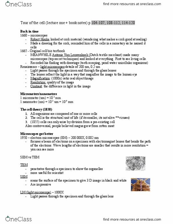
1660 microscopes invented by robert hook but first live specimen saw by. 1838 schleiden and schwann discovered that all organisms are alive and composed of 1 or more cells. Em- electron miscopies: they scan through specimens, there are two types, sem- scanning electron microscope, scans surface creating 3d shape, tem- transmission electron microscope, penetrates right through specimen showing organelles. Two types of cells: prokaryotic, before nucleus, very small cells, ~5um, the small cell is explained by the theory of endosymbiont theory, not compartmentalized, eukaryotic, true nucleus, has organelles, between 10-100 um. *1000 microns (um) = 1mm organelles cell membrane. The cytosol is only the liquid the cytoplasm incudes gritty part and also liquid. The reason it has two membranes is explained by the theory endosymbiont theory. Not an organelle but is in the organelle nucleus. Is made up of dna and protein (histones) The inner and outer membrane of the nucleus.