ANHB2212 Lecture Notes - Lecture 5: Osteoclast, Intramembranous Ossification, Sanguinaria

28 May 2018
School
Course
Professor

L5 Histology: Skeletal Tissues (12 Mar)
Outcomes
1. Cartilage (chondro) 软骨
• 3 types of cartilages
• structure
• function
• growth
2. Bone (os)
• Histological structure
• Mineralization
• Lamellar pattern
• Osteons
• 3 types of Bone cells
• Histological types and Anatomical forms
3. Skeletal tissues = cartilage and bone
• Cartilage and bone are Connective tissues they
have cells and matrix
• In the skeletal tissues- they have matrix, which
are fibers and have spec features of ground
substances (amorphous, jelly like)
4. Cartilage and Bone
• development
• growth
Cartilage (chondro) 软骨
• Root word → Chondro
• Fibrous tissues have various degrees of stretch
resistance
o They can be compressed but has an
extent
▪ due to presence of
glycosaminoglycan (GAG) in the
matrix, -ve charges and presence
of water
o their matrixes have all 3 fibers, ground
substances, special molecules and water etc
• Cartilage has cells → Chondroblasts (active,
cartilage-forming cells) and Chondrocytes
(resting cells)
• Recall: Matrix has all 3 types of fibers, special
molecules & water
3 types of cartilages
1. Hyaline cartilage
• Aka “like glass”
• Has glassy appearance
• Fibres are present but not visible
2. White fibrocartilage
• Has lots of collagen (becomes stretch resistant when
stretched to an extent)
• slightly deformable
3. Elastic cartilage
• Elastic fibres are abundant
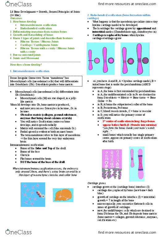
Cartilage, During life
• Cells are in contact with the matrix
• When we process cells for histological section, cells
shrink (right)
• There are “chondrocytes in a lacuna”
o Lacuna = gap
• Lacunae = Characteristic of cartilage and bone
• (left) non-histological specimen
• (right) stained, sectioned histological specimen
1. Hyaline Cartilage (cartilage = soft bone)
• is as Dense Fibrous Connective Tissue
• Avascular
• so, where do they get nutrients from?? Hyaline
cartilage has Perichondrium → it’s the Fibrous
CT that wraps around the cartilage,
o fibrous connective tissue (CT) has blood
vessels (BV), nutrients diffuse through
matrix
• Perichondrium has undifferentiated cells which
are known as Chondroblasts (active)
o Chondroblasts produce new matrix, they
add onto the surface
o Growth mechanism of Perichondrium:
Appositional Growth (explained later),
▪ “growth because of
undifferentiated cells in the
perichondrium”
find more resources at oneclass.com
find more resources at oneclass.com

hyaline cartilage continued
• Chondrocytes revert to Chondroblasts. This is found
Deep inside the cartilage
(Chondroblasts = active) (Chondrocytes = resting)
• Chondrocytes divide and produce new matrix
• When hyaline is viewed under the microscope:
o Cell nests / Isogenous groups are visible
o iso = same, genous = born
o born of the same cell
• Growth mechanism of Chondrocytes:
Interstitial Growth
There are 2 growth mechanisms for Hyaline Cartilage
1. Perichondrium (that has Chondroblasts)– appositional
growth
2. Chondrocytes – Interstitial growth, (form
Chondroblasts)
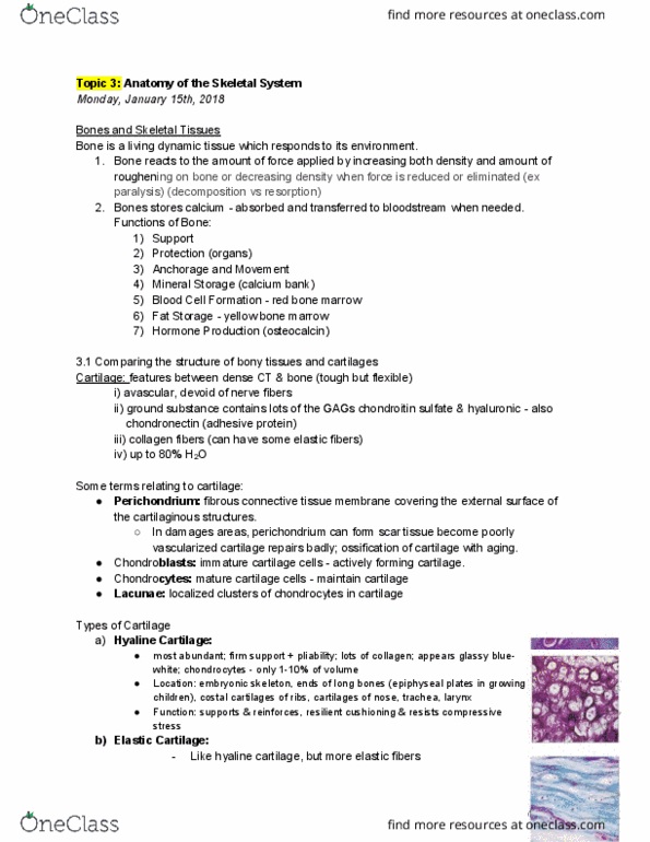
Hyaline cartilage + growth
1. Appositional growth
A- Location of Chondrogenic cells
B- Chondroblasts are newly formed/recruited
This is a mass of hyaline cartilage, everything below the
black line is the hyaline cartilage, H.C is an example of dense
fibrous CT (DICT), fibres are present but not visible
2. Interstitial growth
A- Cells just divided
T- Territorial Matrix
B – 2 cells separate
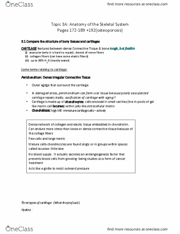
2. White Fibrocartilage
• Predominance of Collagen, is a cartilage that has
collagen
• Has white appearance
• Fibres are visible in sections/ when viewed under
microscope
• “Fibrocartilage is an example of dense FT
→
the
nuclei is not as flat as the FT’ ones. It appears as if
its in a lacuna”
• WFC merges with Fibrous Tissue (FT) around
• WFC has slight flexibility/slightly deformable
• WFC’s cells are in a lacuna, often in rows (there
are bundles of collagen fibres, cells are usually in
rows between collagen fibres)
1. Nuclei are not as flat than the FT’s
2. Absence of blood vessels, cells of white fibrocartilage in a
lacuna
lacuna – an unfilled space, a gap, a cavity or depression in a
bone
find more resources at oneclass.com
find more resources at oneclass.com
Document Summary
3 types of cartilages structure function growth: bone (os, histological structure, mineralization, lamellar pattern, osteons, histological types and anatomical forms. 3 types of bone cells: skeletal tissues = cartilage and bone, cartilage and bone are connective tissues they have cells and matrix. In the skeletal tissues- they have matrix, which are fibers and have spec features of ground substances (amorphous, jelly like: cartilage and bone, development growth. Hyaline cartilage has perichondrium it"s the fibrous. Appositional growth (explained later): growth because of undifferentiated cells in the perichondrium hyaline cartilage continued, white fibrocartilage, chondrocytes revert to chondroblasts. This is found: predominance of collagen, is a cartilage that has. Deep inside the cartilage (chondroblasts = active) (chondrocytes = resting: chondrocytes divide and produce new matrix, when hyaline is viewed under the microscope: Cell nests / isogenous groups are visible iso = same, genous = born: born of the same cell, growth mechanism of chondrocytes: