VETS2011 Lecture Notes - Lecture 4: Macrophage, Internal Jugular Vein, Inferior Vena Cava


CARDIOVASCULAR ANATOMY #3: BLOOD VESSELS
A. OVERVIEW
Arteries + capillaries + veins form continuous transport system lined by endothelium.
*Endothelium: tissues that form single layer of cells lining organs & cavities.
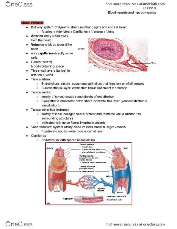
3 basic histological layers @ all blood vessels (vary depending on function of vessel):
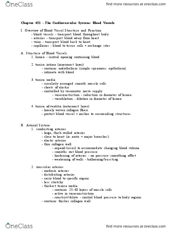
1. Tunica Intima Innermost layer. Consists:
a. Endothelium
squamous cells lining lumen on basement membrane.
prominent nuclei bulging into lumen.
b. Sub-endothelial Coat
fibro-elastic CT
fibroblasts: produces collagen, etc.
myointimal cells: cells of muscle layer of arterial wall; responsible for
organization of deposits @ vessel wall.
All oriented parallel to long axis of vessel.
c. Internal Elastic Lamina
elastic fibres.
2. Tunica Media Middle layer.
circularly-arranged smooth mm. cells
collagen
fibroblast
@ large vessels: nerve (nervi vasorum) + blood supply (vasa vasorum)
3. Tunica Adventitia Outermost layer. Consists:
a. External Elastic Lamina (not always obvious)
b. Dense Fibroelastic Tissue
Blends with surrounding CT.
Nervi vasorum + vasa vasorum ramifies throughout.
1
find more resources at oneclass.com
find more resources at oneclass.com

B. ARTERIES
1
.
Conducting (elastic) Arteries Large | Wide lumen
Function: conduct large vol. of blood around body.
Tunica Intima endothelium
sub-endothelial coat
internal elastic lamina
Tunica Media elastic fibres (predominant)
fine collagen fibres
fibroblasts
smooth mm.
2
find more resources at oneclass.com
find more resources at oneclass.com

Tunica Adventitia CT blends with surrounding tissue.
X distinct external elastic lamina.
2
.
Distributing (muscular) Arteries Small & medium-sized arteries.
Function: carry blood to specific tissues.
thick layer of smooth mm.
Tunica Intima endothelium
sub-endothelial coat
internal elastic lamina
Tunica Media Mainly thick layer of smooth mm.
Tunica Adventitia external elastic lamina
3
.
Arterioles Narrow lumen (< 200μm).
Function: control total peripheral vascular resistance (site
where blood pressure reduced from systemic levels to low
capillary levels).
Tunica Intima endothelium
internal elastic lamina
X sub-endothelial coat.
Tunica Media 1 – 3 layers of smooth mm.
Tunica Adventitia CT blends with surrounding tissue.
X distinct external elastic lamina.
C. CAPILLARIES
Narrow (diameter < 10 μm) – allow passage of single blood cells.
Endothelial cells line basement membrane (surrounded by loose CT).
Function: site of exchange of metabolites + waste products with tissues.
5 types of capillaries:
1. Continuous Capillaries X pores / interruptions between endothelial cells.
Location: muscle, lungs, nervous tissue, brain.
2. Fenestrated Capillaries pores throughout endothelial walls.
Location: site where fluid transport is important (endocrine glands,
intestines, glomerular capillaries of kidney).
3. Sinusoidal Capillaries Larger | Irregular-shaped
X distinct basement membrane.
Location: endocrine organs, aortic body, carotid body.
4. Sinusoids Larger than sinusoidal capillaries.
X basement membrane.
gaps in walls – large molecules (proteins) may be exchanged.
3
find more resources at oneclass.com
find more resources at oneclass.com
Document Summary
Arteries + capillaries + veins form continuous transport system lined by endothelium. *endothelium: tissues that form single layer of cells lining organs & cavities. 3 basic histological layers @ all blood vessels (vary depending on function of vessel): tunica intima. Squamous cells lining lumen on basement membrane. Prominent nuclei bulging into lumen: sub-endothelial coat. Myointimal cells: cells of muscle layer of arterial wall; responsible for organization of deposits @ vessel wall. All oriented parallel to long axis of vessel. @ large vessels: nerve (nervi vasorum) + blood supply (vasa vasorum) Nervi vasorum + vasa vasorum ramifies throughout. Function: conduct large vol. of blood around body. Function: control total peripheral vascular resistance (site where blood pressure reduced from systemic levels to low capillary levels). Narrow (diameter < 10 m) allow passage of single blood cells. Endothelial cells line basement membrane (surrounded by loose ct). Function: site of exchange of metabolites + waste products with tissues.