BIOC2000 Lecture Notes - Lecture 14: Protein Structure, Pymol, Beta Barrel


Lecture 14: An introduction to
protein structure and folding
Illustrating protein structures
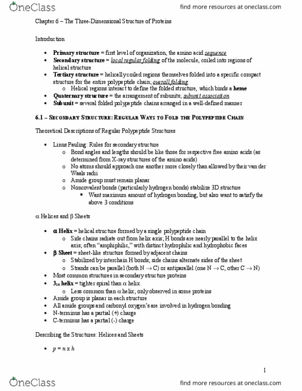
• α helices are shown as ribbons or cylinders
• β strands are shown as arrows, arrow heads always point in the N-
terminus to C-terminus direction
• non / structures are drawn as threads or strings
Protein tertiary structures can be represented using a computer program.
Illustrating protein surfaces
CPK models
In Corey-Pauling Koltun (CPK) models, each atom is represented with a
sphere proportional to the atom’s van der Waal’s (VDW)radius.
- Gives you an idea of the volume taken up by the protein molecule
- However, it doesn’t give you a good idea of the fold of the molecule –
cannot tell if the molecule is made up of helices /sheets
Molecular surface representations
Do not allow easy identification of fold of protein but useful for other
purposes.
There are two surfaces: (*Do not worry too much about)
(a) solvent accessible – surface of protein accessible to solvent
(b) solvent excluded surface (Connelly surface) – region solvent is
excluded from
Advantage of Connelly surface #1 - if you are looking for an interaction
between protein-protein or protein-ligand, you can visualize whether they
have good shape complementarity or not
find more resources at oneclass.com
find more resources at oneclass.com

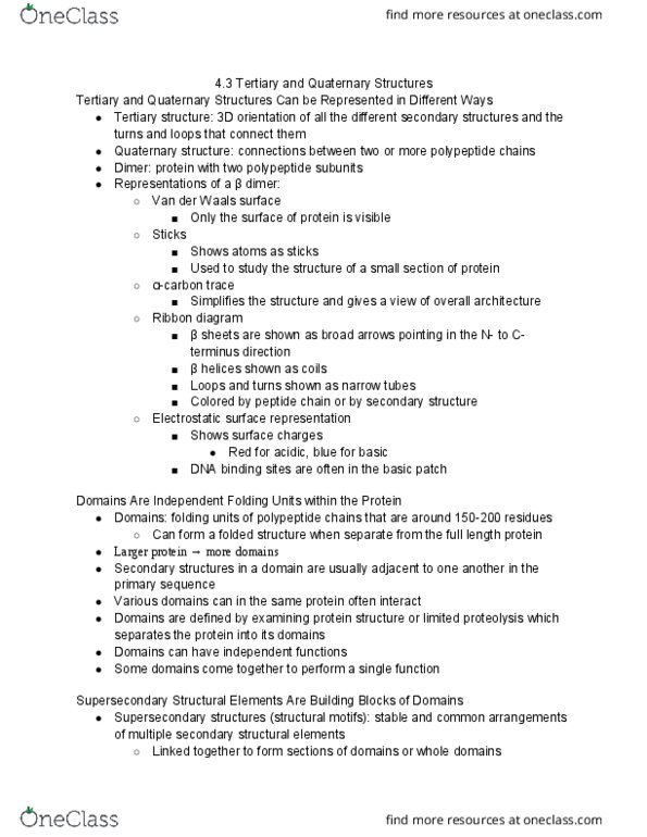
Advantage of Connelly surface #2: Properties of protein can be mapped
onto Connelly surface. Surface is just a representation of the shape of the
molecule, can map what you want onto it.
Common inquiries: Are there parts of the protein highly charged (+ve/-ve)
because that might for example, govern its interaction
The Connelly surface is a good way of visualizing crevices and bumps on
the surface of the protein. It is also ideal for visualizing the electrostatic
potential (charge distribution) on the surface of the protein (blue = +ve,
red = -ve).
This particular toxin has very positively charged surface, and hardly any
negatively charged surface.
Wireframe and ball-and-stick representations
- Similar to CPK but instead of giving atoms their full VDW’s radius, they
are given very smaller radius.
- Important if observing binding of a small molecule and protein-protein
interactions
- Important in drug discovery or drug development for ‘Molecular
docking’ - The goal of ligand-protein docking is to predict the
predominant binding mode(s) of a ligand (your drug) with a protein of
find more resources at oneclass.com
find more resources at oneclass.com
Document Summary
Lecture 14: an introduction to protein structure and folding. Illustrating protein structures: helices are shown as ribbons or cylinders, strands are shown as arrows, arrow heads always point in the n- terminus to c-terminus direction, non (cid:536)/(cid:537) structures are drawn as threads or strings. Protein tertiary structures can be represented using a computer program. In corey-pauling koltun (cpk) models, each atom is represented with a sphere proportional to the atom"s van der waal"s (vdw)radius. Gives you an idea of the volume taken up by the protein molecule. However, it doesn"t give you a good idea of the fold of the molecule cannot tell if the molecule is made up of (cid:536)helices /(cid:537)sheets. Do not allow easy identification of fold of protein but useful for other purposes. There are two surfaces: (*do not worry too much about) (a) solvent accessible surface of protein accessible to solvent (b) solvent excluded surface (connelly surface) region solvent is excluded from.