PHYS20008 Lecture Notes - Lecture 19: Thoracic Wall, Autonomic Nervous System, Bronchiole

12 Jun 2018
School
Department
Course
Professor

Lecture 19
PHYS20008 - HUMAN PHYSIOLOGY
LECTURE 19
RESPIRATORY STRUCTURE & CONTROL
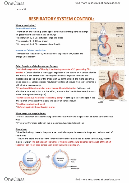
WHAT IS RESPIRATION?
•Ventilation or Breathing: Exchange of air between
atmosphere & lungs.
•Exchange of O2 & CO2 between lungs & blood.
•Transport of O2 & CO2 by blood.
•Exchange of O2 & CO2 between blood & cells.
•At a fundamental level, respiration is basically metabolism.
Always a marker of what’s happening in your body.
INTERNAL OR CELLULAR RESPIRATION
•Intracellular reaction of O2 with nutrients to produce CO2,
water & energy.
RESPIRATORY SYSTEM FUNCTIONS
•Aids in regulation of blood pH by altering amount of H+-
generating CO2 exhaled.
•Provides additional route for water loss & heat elimination
(although not effective in humans).
•Enhances venous return via “respiratory pump”.
•Enables vocalisation & smell.
•Defends against inhaled foreign matter
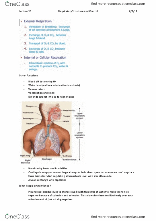
•Ability to speak.
•Difference between upper and lower airways, don’t stress about
it.
BRANCHING OF AIRWAYS
•Bronchioles expand or dilate influence respiration rate.
•Pressure changes determined by pump.
•Rate at which air moves in and out is determined by airways.
•The total cross sectional area is greatest in the alveoli.
•List above. Basically SA. Alveoli SA = tennis court.
STRUCTURE OF RESPIRATORY AIRWAYS
•Trachea and larger bronchi
•Fairly rigid, non-muscular tubes
•Rings of cartilage prevent collapse

Lecture 19
PHYS20008 - HUMAN PHYSIOLOGY
•Bronchioles
•Smooth muscle little tubes leading into airways. Individual are small but have large SA.
•No cartilage to hold them open
•Walls contain smooth muscle innervated by autonomic nervous system to enable them to dilate
during fight/flight response.
•Sensitive to certain hormones and local chemicals
•Alveoli
•Thin-walled sacs
•Site of gas exchange with capillaries
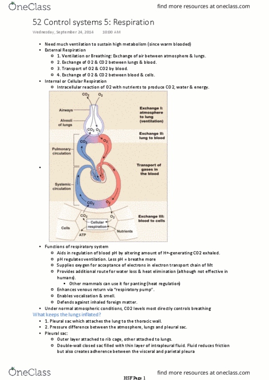
KEEPING THE LUNG INFLATED
•1. Pleural sac which attaches the lung to the thoracic wall.
•2. Pressure difference between the atmosphere, lungs and pleural sac. Always will move from
higher pressure down to lower pressure.
PLEURAL SAC
•The pleural sac holds lungs to wall of ribcage
using only a thin layer of water. It is a double-
wall closed sac filled with intrapleural fluid.
•If the pressure in the pleural sac were higher than
the lungs it would explode the lungs.
PLEURAL SAC & MEMBRANES
•Diagram right.
PRESSURES IMPORTANT IN
VENTILATION
•Diagram below. If the pressure was
equalized the lung would collapse into
itself.
!
Document Summary
Always a marker of what"s happening in your body. Internal or cellular respiration: intracellular reaction of o2 with nutrients to produce co2, water & energy. Branching of airways: bronchioles expand or dilate influence respiration rate, pressure changes determined by pump, rate at which air moves in and out is determined by airways, the total cross sectional area is greatest in the alveoli, list above. Structure of respiratory airways: trachea and larger bronchi, fairly rigid, non-muscular tubes, rings of cartilage prevent collapse. Phys20008 - human physiology: bronchioles, smooth muscle little tubes leading into airways. Pleural sac which attaches the lung to the thoracic wall: 2. Pressure difference between the atmosphere, lungs and pleural sac. Always will move from higher pressure down to lower pressure. Pleural sac: the pleural sac holds lungs to wall of ribcage using only a thin layer of water.