ANAT30008 Lecture Notes - Lecture 7: Referred Pain, Hemiazygos Vein, Recurrent Laryngeal Nerve

17 Aug 2018
School
Department
Course
Professor
Document Summary
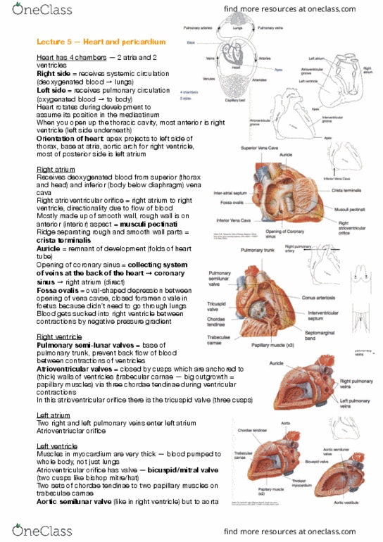
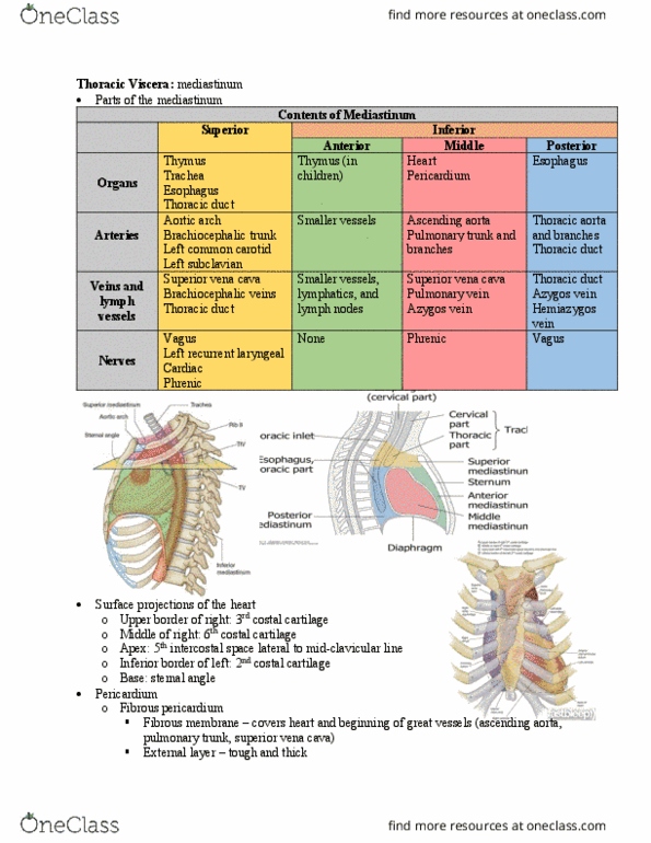
Superior mediastinum (taking away layers from anterior to posterior) Take o manubrium thymus: thymus = development of immune system but becomes redundant after puberty, so shrivels up in elderly people hard to see, looks like attened connective tissue. At the plane of louis, trachea bifurcates into right and left main bronchi (inferior and posterior to pulmonary arteries) Oesophagus behind trachea (and heart) to go through diaphragm. Anterior mediastinum contains fat, lymph nodes, internal thoracic vessels (right up against rib cage) and inferior part of thymus in children. Middle mediastinum contains the heart and the roots of the great vessels + nerves. Posterior mediastinum oesophagus (towards left as descends), descending aorta (towards midline /right from left), thoracic duct and nerves. Note: left vagus nerve runs anterior, right runs posterior. Descending aorta: bronchial arteries to lungs, oesophogeal branches wrap around and supply oesophagus, posterior intercostal arteries for each intercostal space. System comprised of 3 main veins azygos, hemiazygos and accessory azygos vein.