ANAT30007 Lecture Notes - Lecture 25: Knee Bursae, Popliteus Muscle, Vastus Medialis

3 Jun 2018
School
Department
Course
Professor

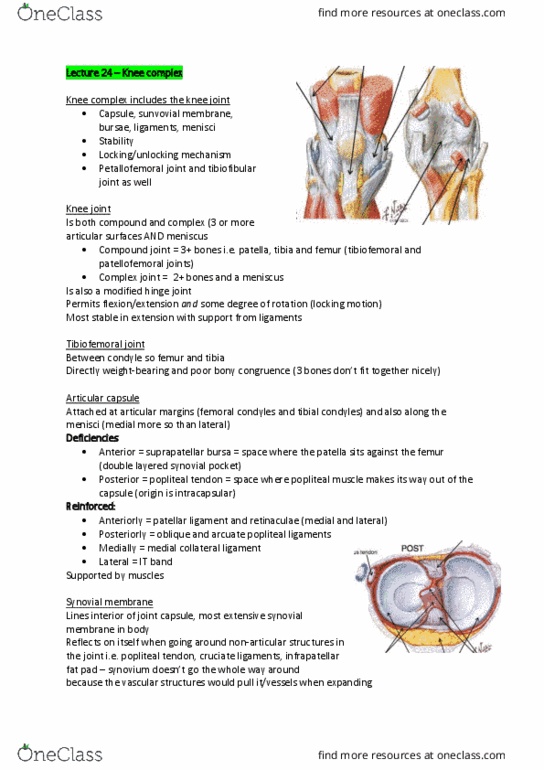
Prevents forward displacement of femur on tibia in WB and backward
displacement of tibia on femur in NWB
○
Prone to injury in flexed knee (bumper car impact)
○
Transverse genicular ligament contacts menisci anteriorly
•
Oblique popliteal ligament
•
Arcuate popliteal ligament
•
Reinforce the capsule, don't have large role in stability
•
Other ligaments
•
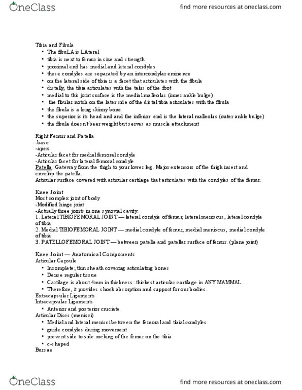
‘C’ shaped fibrocartilagineous wedges: lateral C tighter than medial
•
Improve congruency by increasing contact area by ~1/3 & stabilises knee
•
Bear weight, protect articular surfaces, spread synovial fluid
•
Blood supply on outer rim
•
Menisci move with femur (relative to tibial plateau) in rotation and with tibia in
F/E (relative to femoral condyles)
○
Allow separate movement in joint capsule - 2 separate compartments
•
Lateral meniscus separated from lateral collateral ligament by popliteal tendon
•
Longer, horns (ant/post, strongest point of attachment)are further apart
○
Less mobile due to attachment to MCL
○
Unhappy triad = tear of MCL, MM and ACL due to valgus force/quick
change in direction
▪
Loose body when pinched by MCL
▪
May be ripped or torn:
○
Medial meniscus more commonly injured
•
Removal leads to incidence of osteoarthritis (bone against bone)
•
(Begin as complete discs - centres wear away during development to leave rims)
•
Menisci
Tibiofemoral stability
Locomotor Page 7

•
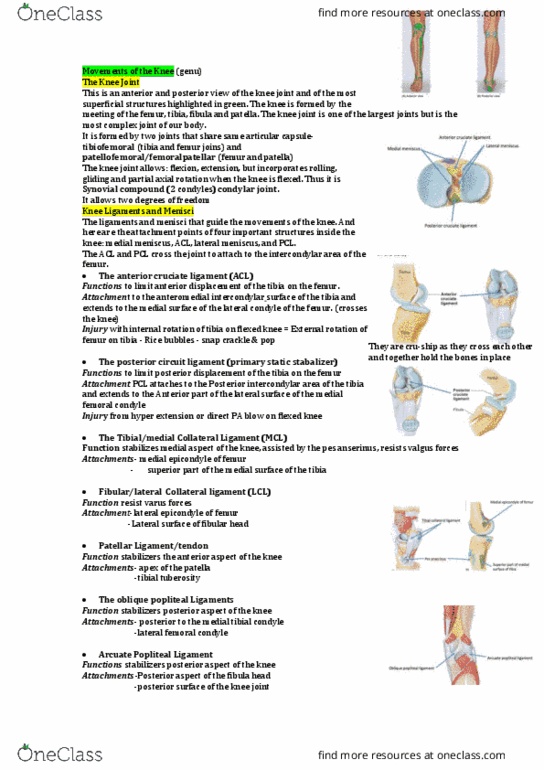
Poor bony congruence
○
Medial condyles longer than lateral (which projects further anteriorly) - creates
locking mechanism
○
Stable in full E, but subject to large forces in WB + twisting
○
Articular surfaces provide little support
•
Cruciate ligaments
○
Collateral ligaments
○
Menisci
○
Stability from 3 pairs of structures:
•
Reinforced by powerful muscles: quadriceps, hamstrings, popliteus, gastrocnemius
•
Maximal stability in full extension (locked): condyles of femur/tibia make full contact
•
Ligament & meniscal damage common in twisting sports - running and turning
•
Complete dislocation uncommon
•
ACL tears and pulls MM/MCL with it
•
Anterior draw test: femur stabilised, tibia moved anteriorly to check for ACL damage if it
moves too easily
•
Posterior draw test: femur stabilised, tibia moved posteriorly to check for PCL damage
•
Knee joint injuries
Muscles and movements
Locomotor Page 8
Document Summary
Prevents forward displacement of femur on tibia in wb and backward displacement of tibia on femur in nwb. Prone to injury in flexed knee (bumper car impact) Reinforce the capsule, don"t have large role in stability. Menisci (cid:858)c(cid:859) shaped fi(cid:271)ro(cid:272)artilagi(cid:374)eous wedges: lateral c tighter tha(cid:374) (cid:373)edial. Improve congruency by increasing contact area by ~1/3 & stabilises knee. Bear weight, protect articular surfaces, spread synovial fluid. Allow separate movement in joint capsule - 2 separate compartments. Menisci move with femur (relative to tibial plateau) in rotation and with tibia in. Lateral meniscus separated from lateral collateral ligament by popliteal tendon. Longer, horns (ant/post, strongest point of attachment)are further apart. Unhappy triad = tear of mcl, mm and acl due to valgus force/quick change in direction. Removal leads to incidence of osteoarthritis (bone against bone) (begin as complete discs - centres wear away during development to leave rims) Medial condyles longer than lateral (which projects further anteriorly) - creates locking mechanism.