ANAT20006 Lecture Notes - Lecture 9: Periodontal Fiber, Hyaline Cartilage, Synovial Joint

23 Jul 2018
School
Department
Course
Professor
Document Summary
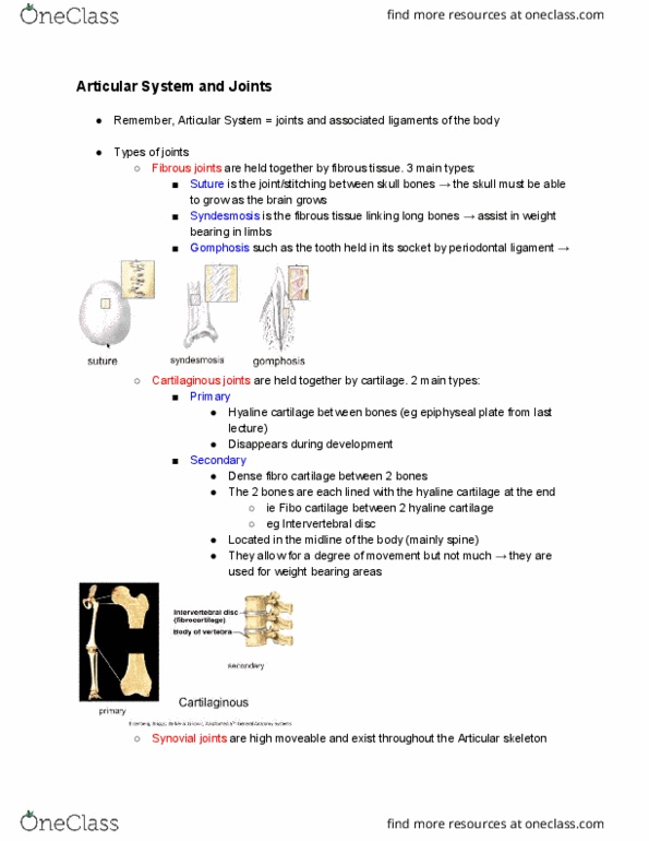
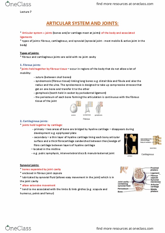
Syndesmosis (fibrous tissue linking long bones eg. distal tibia and fibula) Gomphosis (tooth in socket by periodontal ligament) Hyaline cartilage between bones (disappears during development eg. epiphyseal plate) Wedge of fibro-cartilage between layers layers of hyaline cartilage (located in the midline eg. intervertebral) Pivot (held by ligament in a ring eg. elbow or neck between vertebrae) Saddle (surfaces are both concave and convex) Ball and socket (eg. shoulder for flexion/extension/abduction/adduction/circumduction) Simple: one pair articular surfaces (2 bones meet and wrapped by fibrous capsule) Compound: more than one pair (elbow and knee where femur meets with patella and tibia) Complex: joint cavity subdivided into more than one compartment by. Flexion/extension (sagittal plane divides into left and right) Abduction- from midline/adduction-to midline (coronal plane- anterior/posterior) Rotation (medial and lateral in transverse plane) Muscles can attach and can have openings for communicating structures (tendons/bursa) Sensory nerves of proprioceptive (stretch and joint position) and pain fibres. Poor blood supply so healing is bad.