ANAT20006 Lecture Notes - Lecture 12: Hyaline Cartilage, Lymph Node, Venous Blood

12 Jun 2018
School
Department
Course
Professor

LECTURE 12
VASCULAR SYSTEM & VESSELS
CIRCULATORY SYSTEMS
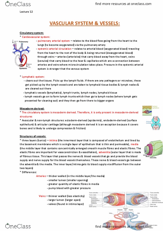
•(1) the cardiovascular system comprises of the pulmonary
arterial system, which takes blood to the lungs for
oxygenation, and the systemic arterial system,when the
oxygenated blood goes around the body. The systemic system
is made up t is made up of arteries and arterioles, veins and
venules and capillaries. Veins take up the deoxygenated blood.
Initially venules, then veins, then superior and inferior vena
cavae.
•The lymphatic system has small lymph vessels. It takes any
fluid, antigens, pathogens etc back to the heart. Lymph tissue
clusters throughout the body. It uses lymph vessels
(lymphatics), lymph trunks, lymph nodes and lymphoid tissue.
•Pulmonary system has lower pressure than systemic.
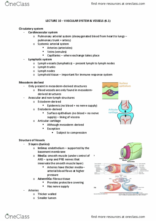
MESODERM DERIVED
•(2) blood vessels are only present in mesoderm derived
structures; not found in epidermis and ectodermal structures.
•Exception: not found in articular cartilage even though it is
mesodermally derived, because it is subject to pressure and
constant rubbing.
•Avascular and non-lymph structures:
•Ectoderm-derived
•Epidermis
•Endoderm-derived
•Surface epithelium
•Articular cartilage
•Although mesoderm derived
•Exception
•Subject to compression
BLOOD VESSEL STRUCTURE
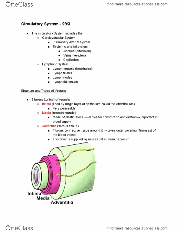
•(3) blood vessels have 3 layers (tunics):
•Intima is connective tissue lined by a single flat layer of endothelium.
•Second layer is media, made up of smooth muscle.
•Adventitia: fibrous tissue. It has the elastic membrane determining how blood vessels contract
and dilate. Outermost layer gets nerve supply. Arteries extend blood out of the heart. Blood is
high pressure when it comes out of the heart. As the blood travels it has to counteract hydrostatic
pressure from gravity, so the pressure in arterial blood is high and the walls are quite thick in
arteries. Systolic pressure is during contraction and diastolic pressure is between contractions.
Arteries having a thicker lumen means they can contract easily.
•Veins are thinner walled. They return deoxygenated blood back to heart. So greater volume and
lower pressure. Veins are assisted by having cusps of valves. Once venous blood goes through a
segment of vein, the flaps close. This is a major difference with arteries; veins have valves.
•Lymphatics also have valves to take up lymph fluid. They travel with
the venous system and go into the heart with the veins.
DIFFERENCES BETWEEN ARTERIES, VEINS AND
LYMPHATICS
•Arteries
Lecture 12 - Friday 18 August 2017
ANAT20006 - HUMAN STRUCTURE & FUNCTION

•Thicker walled
•Smaller lumen
•Elastic fibres in media
•Veins
•Thinner walled
•Larger lumen
•Valves
•Lymphatics
•Thinner walled than veins
•Valves
ARTERIES
TYPES OF ARTERIES
•(4) arteries. The word literally means ‘carry blood’. The heart is very muscular so the arteries
closest to the heart are very elastic. The aorta is very elastic in order to be able to take the change
in pressure. These arteries next to the heart need to be able to expand easily in order to take
pressure off the blood coming through. So these arteries prevent sudden drops in BP.
•Smooth muscle has a strong role in vasoconstriction and vasodilation. Distribution vessels have
the capacity to direct blood to regions of the body in need of blood supply.
•Named according to function: Arterial branches going to bone = nutrient artery. Artery going to
skin = cutaneous arteries. Going to joints = articular arteries.
ELASTIC ARTERIES
•Closest to heart. Have large quantities of elastic tissue and act as
‘conducting’ vessels and prevent sudden drop in blood pressure.
MUSCULAR ARTERIES
•Most named arteries
•Large amount of smooth muscle in media
•Act as ‘distribution’ vessels
•Progressively reducing in calibre
•Redistribution, re-channelling possible
•Arterial branches to somatic structures
Lecture 12 - Friday 18 August 2017
ANAT20006 - HUMAN STRUCTURE & FUNCTION
Document Summary
Circulatory systems: (1) the cardiovascular system comprises of the pulmonary arterial system, which takes blood to the lungs for oxygenation, and the systemic arterial system,when the oxygenated blood goes around the body. The systemic system is made up t is made up of arteries and arterioles, veins and venules and capillaries. Initially venules, then veins, then superior and inferior vena cavae: the lymphatic system has small lymph vessels. It takes any fluid, antigens, pathogens etc back to the heart. It uses lymph vessels (lymphatics), lymph trunks, lymph nodes and lymphoid tissue: pulmonary system has lower pressure than systemic. Blood vessel structure: (3) blood vessels have 3 layers (tunics), intima is connective tissue lined by a single flat layer of endothelium, second layer is media, made up of smooth muscle, adventitia: fibrous tissue. It has the elastic membrane determining how blood vessels contract and dilate. Blood is high pressure when it comes out of the heart.