ANAT20006 Lecture Notes - Lecture 2: Neural Tube Defect, Embryonic Stem Cell, Inner Cell Mass

12 Jun 2018
School
Department
Course
Professor

LECTURE 2
EMBRYOLOGY (1)
SUMMARY OF THIS LECTURE STREAM
•Describe the critical events that take place during embryo development to form a functional
human body
•Understand the cellular basis of the mechanisms of these events
•Use this knowledge to predict what would happen if things do not go to plan
WHY DO WE STUDY EMBRYOLOGY?
•Logical framework for understanding adult anatomy
•Provides information about many issues: reproduction,
contraception, stem cells, etc.
•Informs about when things go wrong: birth
defects, cancer
CONGENITAL DISORDERS
•Congenital disease, birth defect
•Structural (Eg. Polydactyly) or functional (Eg.
metabolic disorder) defects
•Condition present at or before birth (regardless
of cause)
•Genetic, infectious, nutritional and/or
environmental in origin - Occurs in approximately
3% of live birth
•Some examples:
•Orofacial clefts (lip, palate)
1:1000)
•Trisomy 21 (Down) 1:700-900
(1:380 if early termination and
death also counted)
•Heart defects 1:110
•Neural tube defects 1:2400
•Polydactyly 1:1100
•
HUMAN LIFE CYCLE
•Main phase to be discussed is from
the egg to the baby. There are 3
periods in this phase. One of them is
called egg which is technically wrong.
•Diagram right above.
•3 stages:
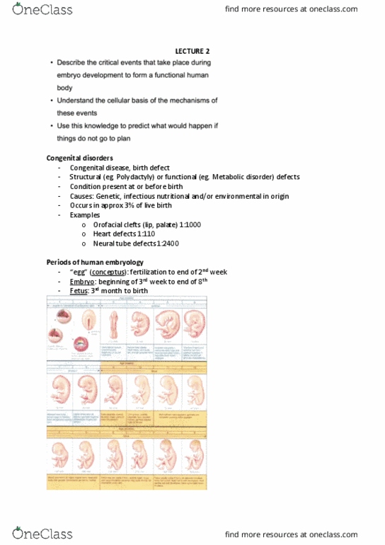
•1. “Egg” (conceptus): fertilization
to end of 2nd week.
•2. Embryo: Beginning of 3rd week
to end of 8th week
•3. Fetus: 3rd month to birth
•The time between stage 1 and 2 is
heavily discussed in terms of things
including abortion etc.
Lecture 2 - Wednesday 26 July 2017
ANAT20006 - HUMAN STRUCTURE & FUNCTION

•Begins with an oocyte being
released from an ovary. The oocyte
has supporting cells to help it to mature. It travels via the
oviduct and meets a sperm to be fertilised. The nuclei of
the oocyte and the sperm fuse to create a zygote. The
zygote begins to divide, the first division giving 2 cells and
the next giving 4 and so on, until you get to a ball of little
cells known as the morula stage. This looks like a
mandarine. These early cell divisions are different to later
cell divisions during embryogenesis and in the adult.
•The early cell divisions create daughter cells that are half the size
of the mother cell whereas later cell divisions also involve cell
growth and will create larger daughter cells.
•Cleavage = smaller and smaller.
Lecture 2 - Wednesday 26 July 2017
ANAT20006 - HUMAN STRUCTURE & FUNCTION

BLASTOCYST STAGE
•Back to the red diagram.
•Morula stage the cells begin to
divide and show morphological
changes. This stage is called the
blastocyst, and we can see 2 types
of cells plus a cavity forming so
there are cells around the cavity
and cells in the middle. Like a
balloon containing cells and also
with cells around the outside.
•Two types of cell
•Outer epithelial layer
(trophoblast)
•Inner cell mass or embryonic stem cells (same thing).
These give rise to the embryo.
•Trophoblast forms extraembryonic structures (part of
placenta)
•Between 5 and 10 days, blastocyst implants into uterine
wall
•DON’T NEED TO KNOW TIMES OF SHIT
HAPPENING.
•Moves down th oviduct into the uterus and then
implants into the uterine wall. The trophoblasts sit
inside the uterine wall. The inner cell mass keeps
differentiating these cells.
TWO GERM LAYER STAGE
•Inner cell mass splits
•Cavities form
•Forming embryonic disc
•Blue cells: epiblasts
•Yellow cells: hypoblasts
•Cavities form within both blue and yellow cell masses.
So the overall balloon is divided into 2 cavities.
•Embryonic disc gives rise to the embryo
Lecture 2 - Wednesday 26 July 2017
ANAT20006 - HUMAN STRUCTURE & FUNCTION
Document Summary
Why do we study embryology: logical framework for understanding adult anatomy, provides information about many issues: reproduction, contraception, stem cells, etc, informs about when things go wrong: birth defects, cancer. Polydactyly) or functional (eg. metabolic disorder) defects: condition present at or before birth (regardless of cause, genetic, infectious, nutritional and/or environmental in origin - occurs in approximately. 3% of live birth: some examples, orofacial clefts (lip, palate) 1:1000: trisomy 21 (down) 1:700-900 (1:380 if early termination and death also counted, heart defects 1:110, neural tube defects 1:2400, polydactyly 1:1100. Human life cycle: main phase to be discussed is from the egg to the baby. One of them is called egg which is technically wrong: diagram right above, 3 stages, 1. (cid:284)egg(cid:285) (conceptus): fertilization to end of 2nd week, 2. Embryo: beginning of 3rd week to end of 8th week: 3.