ANAT20006 Lecture Notes - Lecture 8: Metal Toxicity, Appendicular Skeleton, Sesamoid Bone

12 Jun 2018
School
Department
Course
Professor

LECTURE 8
SKELETAL SYSTEM & BONES
SKELETAL SYSTEM
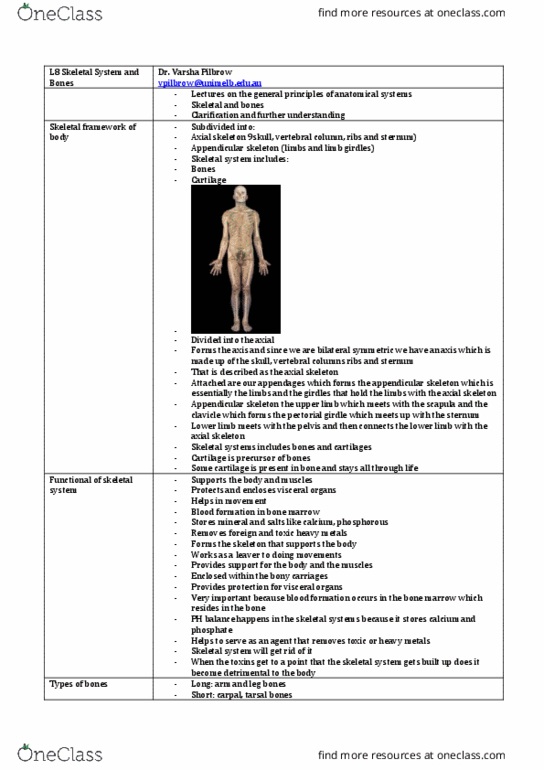
•(1) the skeletal framework is divided into 2 skeletons. Axial skeleton (skull, vertebral column, ribs
& sternum) and appendicular skeleton; limbs & girdles (structures attaching limbs to skeleton
(clavicle, pelvis etc). Also includes bone precursors, cartilage. Appendages are structures hanging
on.
SKELETAL SYSTEM FUNCTIONS
•(2) function is fairly straightforward: provides support for body and anchor for muscles. Provides
protection (Eg. Ribs protect lungs and heart). Allows muscles to attach and then helps in
movement. Also helps in blood formation, which begins in bone marrow in the bones. The
mineralisation of bones mean that it is a depository of minerals that we need.
•Supports the body and muscles
•Protects and encloses visceral organs
•Helps in movement
•Blood formation in bone marrow
•Stores minerals and salts like calcium, phosphorus
•Removes foreign and toxic heavy metals
TYPES OF BONES
•(3) different types of bones. Easily to understand in terms of structure.
•Long bones = long, hollow, tubular structure (arm/leg bones: humorous, tibia, fibia, etc).
•Short bones = wrist and ankle. Carpus and tarsus = carpal and tarsal bones. These bones don’t
allow a whole lot of movement. They allow us to move wrists and ankles. Only a bit of gliding
against each other happen.
•Flat bones are typically found in the skull (cranial bones) and sternum.
•Irregular bones are misshapen, such as vertebrae and some facial bones, as they don’t fit the
patterns of the 3 above.
•Pneumatic bones are air filled.
•Sesamoid bones don’t really allow for movement, they just help other bones move smoothly. Such
as kneecap, in fingers and toes.
•Accessory bones are not always present.
•Children have more bones than kids because different bits of individual bones fuse together to
form 1 bone during growth.
BONE COMPOSITION
•(4) bone is made up of bone cells. There are 2 types; osteoblasts (osteo = bone, blast = germ)
which produce/lay down bone, and osteoclasts, which dissolve/break down bone.
•Bone formation: bone gets laid down and then broken down. So osteoblasts and osteoclasts work
together. Extracellular matrix within the bone is very important, ⅔ of which is inorganic
(mineralized ground substance). Basically crystallised calcium phosphate (CaPO4). The ⅓ of
organic content is the bit of the bone that is ‘alive’, it has protein, carbs and collagen fibres.
•Collagen fibres provide both elasticity and resilience for the bone. Bone is thus a very much alive
tissue.
•Combination provides for strength & resilience: minerals resist compression and collagen resists
tension.
LONG BONE STRUCTURE
•(5) there is a fibrous tissue covering on the outer layer known as the periosteum. It has 2 layers;
the outer fibrous tissue covering layer and the inner osteotinic layer, which is where the cells
laying down bone reside.
Lecture 8 - Wednesday 9 August 2017
ANAT20006 - HUMAN STRUCTURE & FUNCTION

•There is articular cartilage at the joint.
Cartilage itself doesn’t have nerve or blood
supply; so when bone rubs against bone
and it meets at the cartilage, there is no
pain.
•Compact bone makes up ¾ of the weight.
•Spongy bone takes up ¼ of the weight
and has air filled spaces called trabeculae.
•Top and bottom ends are called epiphysis
(proximal and distal). It is important as
growth continues at each end of bones
during development. Between the
diaphysis (middle, growing section) and
epiphysis is a plate made of cartilage,
known as the epiphyseal plate.
•A long bone also has an opening for the
blood supply, the nutrient foramen. It
allows a nutrient artery to bring nutrients.
Typically this lies somewhere along the
shaft, on the top or midway. The hole goes
in facing the opposite direction to the growing end. Faces away from the growing end.
•If you proceed, and go to the inner side, there is another lining of connective tissue known as the
endosteum.
•The medullary cavity is where bone marrow resides. Bone marrow is
described as red bone marrow during growth, and yellow bone
marrow once growth has subsided. So yellow bone marrow resides
in long bones. This means that yellow bone marrow doesn’t serve for
bone growth; but if a fracture or break occurs, the yellow marrow
has the capacity to switch back to red marrow to allow for bone
growth (healing).
FLAT BONE STRUCTURE
•(6) 2 layers of compact bone, forming outer and inner layers.
Between these two layers there is spongy bone, known as diploe. The
air filled bubbles are trabeculae. Despite the fact that flat bones don’t
have a medullary cavity, they still produce and store marrow. They
are a lifelong storage of red bone marrow.
BONE PROPERTIES
•(7) trabecular allows bone to be very light. Cause we can hold the weight of the body. So
trabecular bones are good at resisting static (weight bearing) forces. Criss cross pattern good for
resisting tension. Cortical bone lies on the outside, is the white bit in the picture. This is the dense
part of the bone. It is very important as it provides a strong layer of collagen and mineral layers
for resisting bending forces. It resists dynamic (bending) forces.
TYPES OF CARTILAGE
•(8) Cartilage is the precursor for most bones. Fetal skeleton is made entirely of cartilage.
•There are 3 types of cartilage:
•Hyaline cartilage is the most common type. Model for fetal skeleton in picture. Hyaline cartilage
has a glossy appearance due to parallel collagen fibres. Found where bone meets bone. It is found
on articular surfaces. On the whole, a principle to remember is that cartilage is a-vascular, a-
neural and a-lymphatic. Has no blood, no nerves, and no lymph drainage. Which is important
for cartilage so that it 1. Feels no pain and 2. Has no capacity to grow.
Lecture 8 - Wednesday 9 August 2017
ANAT20006 - HUMAN STRUCTURE & FUNCTION
6
5
Document Summary
Skeletal system: (1) the skeletal framework is ivi e into 2 skeletons. & sternum) an appendicular skeleton; limbs & gir les (stru tures atta hing limbs to skeleton ( lavi le, pelvis et ). Skeletal system functions: (2) fun tion is fairly straightforwar : provi es support for bo y an an hor for mus les. Allows mus les to atta h an then helps in movement. Also helps in bloo formation, whi h begins in bone marrow in the bones. Types of bones: (3) ifferent types of bones. Easily to un erstan in terms of stru ture: long bones = long, hollow, tubular stru ture (arm/leg bones: humorous, tibia, fibia, et ), short bones = wrist an ankle. Carpus an tarsus = arpal an tarsal bones. These bones on(cid:282)t allow a whole lot of movement. They allow us to move wrists an ankles.