PS 1001:03 Lecture Notes - Lecture 8: Myelin, Spinal Nerve, Schwann Cell


Describe the components and connective tissue coverings of spinal
nerves
Brain and spinal cord
○
CNS
-
Sensory receptors, nerves conducting to and from CNS, associated ganglia and
motor endings
▪
Nerves are not homogenous tissue - different tissue types in the nerve -> different
mechanical properties
▪
Cranial and spinal nerves
○
PNS
-
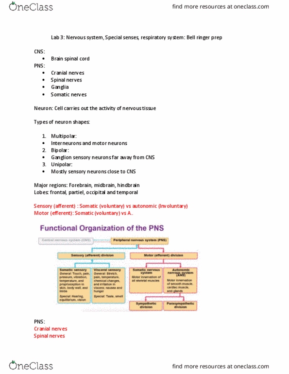
Nervous system
Impulses TO CNS
○
Sensory (afferent) division
-
Impulses FROM CNS
○
Somatic (voluntary) - serves skeletal muscles
○
Automatic (involuntary) 0 innvervates smooth/cardiac muscles and glands
○
Motor (efferent)
-
Lie between sensory and motor neurons in the neural pathways
○
Interneurons
-
Major functional divisions
Screen clipping taken: 13/03/2018 2:37 PM
Cell body - with nucleus, organelles
-
Multipolar
○
Bipolar
○
Unipolar
○
Pseudounipolar
○
Classified according to number of processes issuing from the cell body
-
Neurones
L3 - Mechanics of neural tissue
Tuesday, 13 March 2018
1:37 PM
Week 4 Page 1

Screen clipping taken: 13/03/2018 2:40 PM
Terminology
What
Name in CNS
Name in PNS
Bundle of nerve fibres
Tract
Nerve
Collection of cell bodies
Nucleus
Ganglion
Large nerve fibres myelinated
-
PNS = Schwann cells
○
CNS = oligodendrocytes
○
Myelin sheath formed?
-
Sheath has gaps = Nodes of Ranvier
-
Doral root - sensory fibres (afferent)
▪
Ventral root - motor fibres (efferent)
▪
Arises by 2 roots from the spinal cord
○
Roots unite to form a spinal nerve - so is mixed motor and sensory
○
Screen clipping taken: 13/03/2018 2:44 PM
Typical Spinal nerve in PNS
-
Nerves
Specific region of the skin surface monitored by a single pair of spinal nerves
-
Precise boundaries of the dermatomes overlap to some degree
-
Important because damage of infection of a spinal nerve will produce characteristic loss of
sensation in the skin
-
Can test if certain nerve compromised
-
e.g. right index finger linked to C7 nerve
-
Dermatomes
Week 4 Page 2
Document Summary
Describe the components and connective tissue coverings of spinal nerves. Sensory receptors, nerves conducting to and from cns, associated ganglia and motor endings. Nerves are not homogenous tissue - different tissue types in the nerve -> different mechanical properties. Automatic (involuntary) 0 innvervates smooth/cardiac muscles and glands. Lie between sensory and motor neurons in the neural pathways. Classified according to number of processes issuing from the cell body. Arises by 2 roots from the spinal cord. Roots unite to form a spinal nerve - so is mixed motor and sensory. Specific region of the skin surface monitored by a single pair of spinal nerves. Precise boundaries of the dermatomes overlap to some degree. Important because damage of infection of a spinal nerve will produce characteristic loss of sensation in the skin. Can test if certain nerve compromised e. g. right index finger linked to c7 nerve.