MMED1005 Lecture Notes - Lecture 12: Ataxia, Simultanagnosia, Thalamus

7 Jun 2018
School
Department
Course
Professor

Main points:
- The retina, photoreceptors and function of other retinal cells
- Transduction of light in photoreceptors
- Disorder of photoreceptors
- Transmission of visual signal from the eye to the brain
- Processing of visual signal in the brain
- Disorders of visual processing
find more resources at oneclass.com
find more resources at oneclass.com

‘eisio fro esterdas leture:
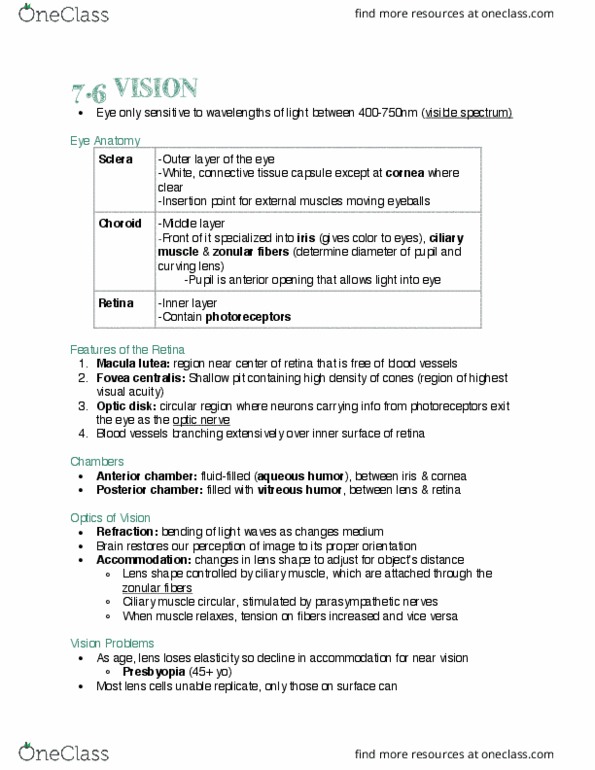
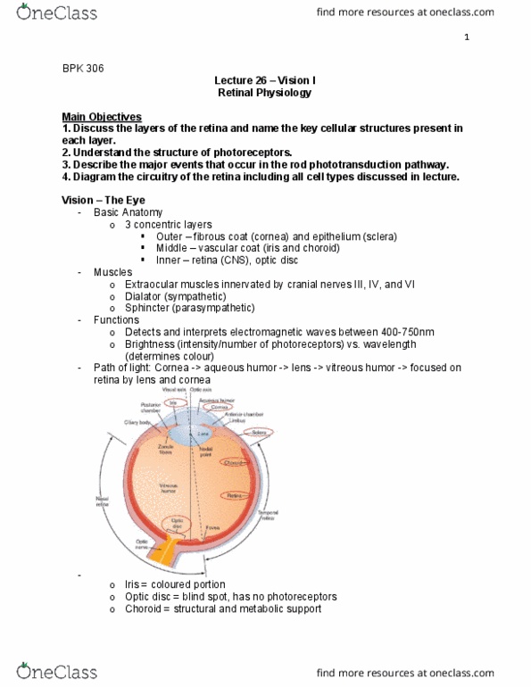
Light enter the eye from the cornea (the clear part), then the aqueous the humor, the lens,
the vitreous humor, the retina,
Cellular organisation of the retina: (two main parts)
- Pigmented epithelium layer:
o nourishes the photoreceptor cells
o is firmly attached to the underlying choroid and
overlying retinal photoreceptor cells
-
- Neuro-retina: Three main neuronal layers of the retina:
o Photoreceptors (rods and cones) – light sensing cells – outermost
layer
o Bipolar cells (span the entire thickness of the retina) – middle layer
o Ganglion cells (their axons join to become the optic nerve) –
innermost layer
- The retina also contains two other types of neurons – horizontal cells and
amacrine cells
Optic nerve: (and the posterior aspect of the eyeball)
- Axons of the ganglion cells (make a right angle turn and) form the optic nerve that
leaves the back of the eyeball at the optic disk
- The exit point of the axons, the optic disk, is the blind spot
(because there are only axons here, no neurons)
find more resources at oneclass.com
find more resources at oneclass.com

Transmission of visual responses in the retina:
- The signals produced in the photoreceptors in response to
light are transmitted to the bipolar cells, then to the ganglion
cells, where action potentials are generated
- These action potentials are carried by the optic nerve to the
brain
The photoreceptor cells:
- Two types of photoreceptors:
o Cones: three types (blue, green, red) depending on the wavelength of light
they absorb, short and thick
o Rods: thin and slender
- In the human retina, rods:cones = 20:1, distriutio ot uifor esterdas le
Excitation of rods and cones:
- Rods and cones contain unique photo-pigments(proteins that absorb light) and thus
absorb different wavelengths of visible light and have different thresholds of
activation
- Rods:
o Very sensitive, respond to very dim light, suited to night vision and peripheral
vision
o Inputs are perceived in grey tones, provide contrast sensitivity
- Cones:
o Low sensitivity, need bright light for activation, suited for light vision
o Each type of cones absorbs different wavelength, provide colour vision
Neural connectivity of photoreceptors
- Rods and cones are connected differently to bipolar and ganglion cells
- Rods:
o Participate in converging pathways – multiple rods feed their input to a single
ganglion cell
o Results in fuzzy vision due to poor resolution
- Cones:
o Each cone in the fovea feeds into a single ganglion cell
o Results in clear vision due to high resolution
- This is why the fovea (cone-rich) provides detailed (central) vision, whereas the
peripheral retina (rod-rich, provides peripheral vision) does not
find more resources at oneclass.com
find more resources at oneclass.com
Document Summary
The retina, photoreceptors and function of other retinal cells. Transmission of visual signal from the eye to the brain. Processing of visual signal in the brain. Light enter the eye from the cornea (the clear part), then the aqueous the humor, the lens, the vitreous humor, the retina, Cellular organisation of the retina: (two main parts) Pigmented epithelium layer: nourishes the photoreceptor cells is firmly attached to the underlying choroid and overlying retinal photoreceptor cells. The retina also contains two other types of neurons horizontal cells and amacrine cells. Optic nerve: (and the posterior aspect of the eyeball) Axons of the ganglion cells (make a right angle turn and) form the optic nerve that leaves the back of the eyeball at the optic disk. The exit point of the axons, the optic disk, is the blind spot (because there are only axons here, no neurons)