HUMB1000 Lecture Notes - Lecture 18: Pulmonary Vein, Simple Squamous Epithelium, Atrioventricular Node

Document Summary
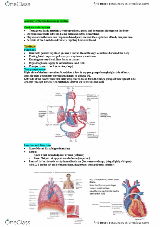
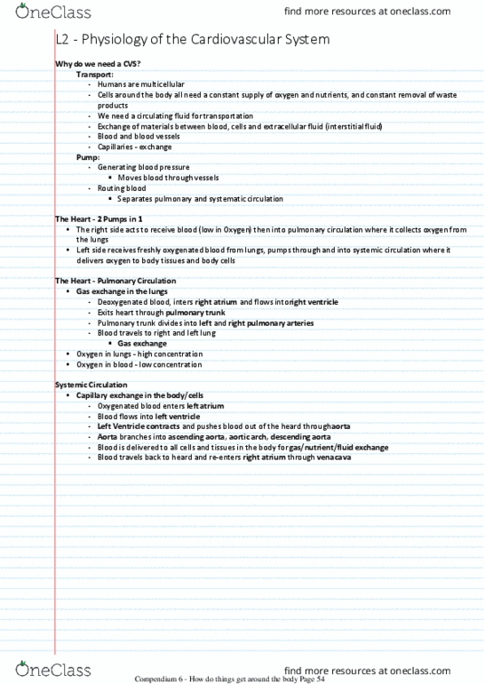
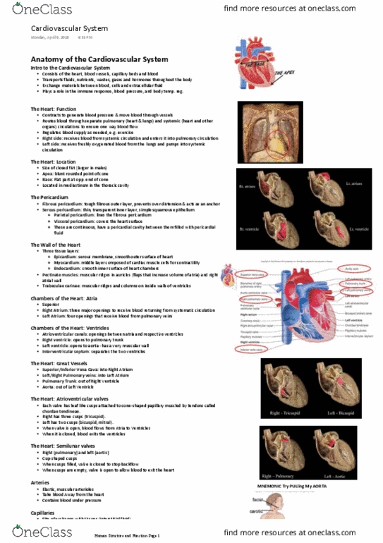
The heart - functions: generating blood pressure. Moves blood through vessels: routing blood. Separates pulmonary and systematic circulations: ensuring one-way blood flow, regulating blood supply, changes to match need. The heart - location: size of a closed fist. Generally larger in males than females: shape. Flat part at opposite of end of cone: located in thoracic cavity in mediastinum, 2/3 of the heart is on the left side. This is why the left lung is smaller than the right lung. Thin, transparent, inner layer of simple squamous epithelium. The two are continuous and have pericardial cavity between them filled with pericardial fluid. The heart - wall: three layers of tissue, epicardium. Serous membrane; smooth outer surface of heart: myocardium. Middle layer composed of cardiac muscle cells - contractility. Compendium 6 - how do things get around the body page 50. Muscular ridges in auricles and right atrial wall: trabeculae carnae. Muscular ridges and columns on inside walls of ventricles.