BIOL125 Lecture Notes - Lecture 8: Sine Wave, Tunica Media, Varicose Veins


BIOL week 8 LC Anatomy of the Vascular System
The CVS: Blood Vessels
- The blood vessels of the body form a closed delivery system that begins and ends at
the heart (blood cannot escape – remains in the circulation)
- Blood vessels transport hormones, oxygen (as haemoglobin), carbon dioxide (as
bicarbonate ions)
- Vena cava is largest vein – transports blood back to the heart and then to the lungs
- Functions:
oDistribution of blood
oExchange of materials at the capillaries
oReturn of blood to the heart
Types of Blood Vessels
- Arteries carry blood away from heart; large amount of smooth muscle to maintain
pressure; branch repeatedly into smaller arteries then into arterioles
- Arterioles carry blood to capillary beds; contain smooth muscle to maintain
pressure
- Capillaries site of diffusion of gases, nutrients and wastes between blood and
interstitial fluid (ISF); leads into venules; have only 1 layer of endothelia cells
- Venules larger veins direct blood into small veins
- Veins direct blood back to the heart; join together to flow into superior and inferior
vena cava
- Arteries and veins supplying same region lie side by side eg. Femoral artery and vein
- The nervous system, through vasoconstriction and vasodilation, can control smooth
muscle in blood vessel wall
- Aorta transports blood from the left ventricle contains largest amount of elastin
fibres, allowing contraction
find more resources at oneclass.com
find more resources at oneclass.com

- Fenestrated capillary have pores through which more materials can come through
– more permeable than continuous capillary
- Sinusoid capillary has most permeability due to larger pores
- Tunica media is where blood innervates artery; responsible for lumen size
- Function of tuncia externa is to support and protect
- Walls of the arteries are much thicker
- Lumen size is wider in the veins
- Arteries can expand and recoil passively
- High pressure system for arteries and low pressure system for veins
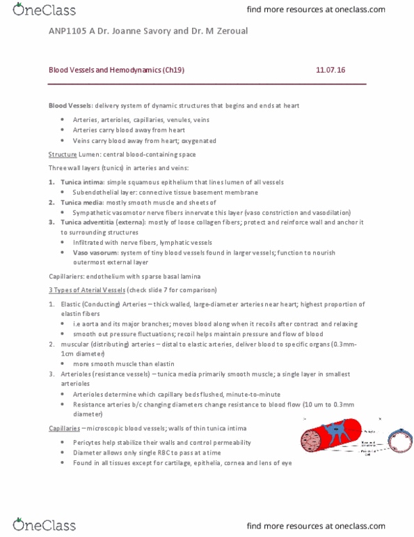
Structure of Blood Vessel Walls
- Walls of arteries and veins have three layers: tunica intima (inner), tunica media
(middle), and tunica externa (outer)
- Tunica intima endothelium lining and surrounding connective tissue with variable
number of elastic fibres; arteries also have thick internal elastic membrane in outer
part
- Tunica media thickest layer in most arteries; concentric smooth muscle sheets in
loose connective tissue framework; has thin band of elastic fibres beneath externa
layer ie. External elastic membrane
- Tunica externa connective tissue sheath; in arteries has collagen fibres and
scattered bands of elastin fibres; generally thickest layer in veins; anchors blood
vessel into surrounding tissue
Differences between arteries and veins
- Vessel wall walls of arteries are thicker than those of veins. Tunica media of
arteries has more smooth muscle and elastic fibres to resist arterial pressure as
ventricles pump blood
- Vessel lumen when not opposed by blood pressure (BP), elastic fibres in arterial
walls recoil reducing size of lumen; lumen keeps round shape but can narrow due
vasoconstriction and vasodilation, controlling blood flow through circulatory system.
Veins are capacitance vessels (can expand to allow more blood through); veins have
wide lumens
- Vessel lining arterial endothelium cannot contract – pleated/folded appearance in
constricted artery. Veins do not have folds (contraction rarely required in venous
tissue)
In the veins, blood flow is assisted by the function of skeletal muscles - valves
- Arteries retain their cylindrical shape, but veins may collapse
- Arteries are more resilient: may be stretched out of shape then return to normal
- Veins cannot tolerate much distortion before tearing or collapsing
- Amount of elastic fibres, collagen and smooth muscle is greater in arteries than in
veins
find more resources at oneclass.com
find more resources at oneclass.com
Document Summary
Biol week 8 lc anatomy of the vascular system. The blood vessels of the body form a closed delivery system that begins and ends at the heart (blood cannot escape remains in the circulation) Blood vessels transport hormones, oxygen (as haemoglobin), carbon dioxide (as bicarbonate ions) Vena cava is largest vein transports blood back to the heart and then to the lungs. Functions: distribution of blood, exchange of materials at the capillaries, return of blood to the heart. Arteries carry blood away from heart; large amount of smooth muscle to maintain pressure; branch repeatedly into smaller arteries then into arterioles. Arterioles carry blood to capillary beds; contain smooth muscle to maintain pressure. Capillaries site of diffusion of gases, nutrients and wastes between blood and interstitial fluid (isf); leads into venules; have only 1 layer of endothelia cells. Venules larger veins direct blood into small veins.