MCB 3020C Chapter Notes - Chapter 1: Scanning Electron Microscope, Antigen, Bright-Field Microscopy

Document Summary
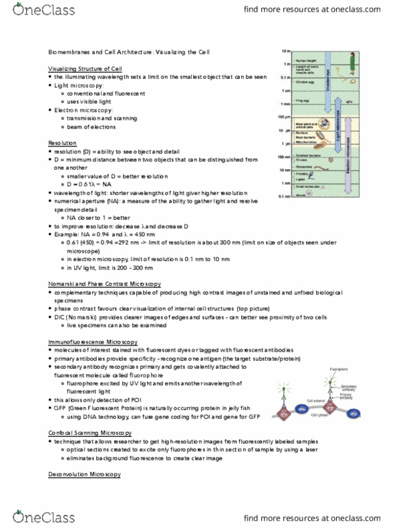
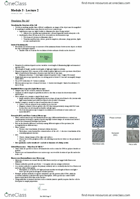
The light microscope allowed the identification of cellular organelles: source of visible light, system of lenses. Limit of resolution: how far apart objects must be to appear as separate points, related to the wavelength of light used for illumination, typical limit of resolution for a light microscope = 200nm, maximum magnification 1000-1400x. Bright field microscopy: while light passed through a specimen, stained or unstrained, most specimens must be sectioned and stained to be made visible, fixed (preserved, not for live specimens. Fluorescence microscopy: allows the detection of specific proteins, live or preserved sample (depending on how samples are prepared, live: e. g. , genetic labeling using green fluorescent protein (gfp, fixed: florescent dyes or antibody staining. Immune system protein: binds to a particular target protein (antigen) with high specifity, fluorescent dyes can be covalently attached to antibodies. Only one antibody used: labeled with fluorescent dye, binds to target protein (antigen) in the sample.